 Movie
Movie Controller


+ Open data

- Basic information
| Entry |  Database: PDB chemical components / ID: HL0 Database: PDB chemical components / ID: HL0 |
|---|---|
| Name | Name: |
-Chemical information
| Composition |  | ||||||
|---|---|---|---|---|---|---|---|
| Others | Type: NON-POLYMER / PDB classification: HETAIN / Three letter code: HL0 / Ideal coordinates details: Corina / Model coordinates PDB-ID: 3QP4 | ||||||
| History |
| ||||||
 External links External links | UniChem / ChemSpider / BindingDB / Brenda / ChEBI / ChEMBL / CompTox / LipidMaps / PubChem / SureChEMBL / Wikipedia search / Google search |
- Structure visualization
| Structure viewer | Molecule:  MolmilJmol/JSmol MolmilJmol/JSmol |
|---|
-Details
-SMILES
| ACDLabs 12.01 | | CACTVS 3.370 | OpenEye OEToolkits 1.7.0 | |
|---|
-SMILES CANONICAL
| CACTVS 3.370 | | OpenEye OEToolkits 1.7.0 | |
|---|
-InChI
| InChI 1.03 |
|---|
-InChIKey
| InChI 1.03 |
|---|
-SYSTEMATIC NAME
| ACDLabs 12.01 | | OpenEye OEToolkits 1.7.0 | |
|---|
-PDB entries
Showing all 5 items
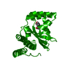
PDB-3qp4: 
Crystal structure of CviR ligand-binding domain bound to C10-HSL
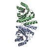
PDB-3qp8:
Crystal structure of CviR (Chromobacterium violaceum 12472) ligand-binding domain bound to C10-HSL

PDB-9iss:
Crystal Structure of Cytochrome P450BM3 III-10C1 Mutant Heme Domain with N-Decanoyl-L-Homoserine Lactone

PDB-9ist:
Crystal Structure of Cytochrome P450BM3 VI-18A12 Mutant Heme Domain with N-Decanoyl-L-Homoserine Lactone

PDB-9isu:
Crystal Structure of Cytochrome P450BM3 V-19A14 Mutant Heme Domain with N-Decanoyl-L-Homoserine Lactone
