 Movie
Movie Controller

[English] 日本語
 Yorodumi
- ChemComp-BAU: 1-((2-HYDROXYETHOXY)METHYL)-5-BENZYLPYRIMIDINE-2,4(1H,3H)-DIONE -
+ Open data

- Basic information
| Entry | 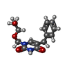 Database: PDB chemical components / ID: BAU Database: PDB chemical components / ID: BAU |
|---|---|
| Name | Name: |
-Chemical information
| Composition |  | ||||
|---|---|---|---|---|---|
| Others | Type: NON-POLYMER / PDB classification: HETAIN / Three letter code: BAU / Model coordinates PDB-ID: 1U1C | ||||
| History |
| ||||
 External links External links | UniChem / ChemSpider / BindingDB / Brenda / ChEBI / ChEMBL / CompTox / DrugBank / HMDB / PubChem / SureChEMBL / Wikipedia search / Google search |
- Structure visualization
| Structure viewer | Molecule:  MolmilJmol/JSmol MolmilJmol/JSmol |
|---|
-Details
-SMILES
| ACDLabs 10.04 | | CACTVS 3.341 | OpenEye OEToolkits 1.5.0 | |
|---|
-SMILES CANONICAL
| CACTVS 3.341 | | OpenEye OEToolkits 1.5.0 | |
|---|
-InChI
| InChI 1.03 |
|---|
-InChIKey
| InChI 1.03 |
|---|
-SYSTEMATIC NAME
| ACDLabs 10.04 | | OpenEye OEToolkits 1.5.0 | |
|---|
-PDB entries
Showing all 4 items

PDB-1u1c: 
Structure of E. coli uridine phosphorylase complexed to 5-benzylacyclouridine (BAU)

PDB-3euf:
Crystal structure of BAU-bound human uridine phosphorylase 1

PDB-3p0e:
Structure of hUPP2 in an active conformation with bound 5-benzylacyclouridine

PDB-3p0f:
Structure of hUPP2 in an inactive conformation with bound 5-benzylacyclouridine
