 Movie
Movie Controller


+ Open data

- Basic information
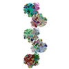
| Entry | Database: PDB / ID: 1h1k | ||||||
|---|---|---|---|---|---|---|---|
| Title | THE BLUETONGUE VIRUS (BTV) CORE BINDS DSRNA | ||||||
 Components Components | (RNA) x 6 | ||||||
 Keywords Keywords | VIRUS/VIRAL PROTEIN/RNA / VIRUS / VIRAL PROTEIN / RNA / VIRUS-VIRAL PROTEIN-RNA complex | ||||||
| Function / homology | RNA / RNA (> 10) / RNA (> 100) Function and homology information Function and homology information | ||||||
| Biological species |  BLUETONGUE VIRUS BLUETONGUE VIRUS | ||||||
| Method |  X-RAY DIFFRACTION / SYNCHROTRON / MOLECULAR REPLACEMENT / Resolution: 10 Å X-RAY DIFFRACTION / SYNCHROTRON / MOLECULAR REPLACEMENT / Resolution: 10 Å | ||||||
 Authors Authors | Diprose, J.M. / Grimes, J.M. / Sutton, G.C. / Burroughs, J.N. / Meyer, A. / Maan, S. / Mertens, P.P.C. / Stuart, D.I. | ||||||
 Citation Citation | Journal: J.Virol. / Year: 2002 Title: The Core of Bluetongue Virus Binds Double-Stranded RNA Authors: Diprose, J.M. / Grimes, J.M. / Sutton, G.C. / Burroughs, J.N. / Meyer, A. / Maan, S. / Mertens, P.P.C. / Stuart, D.I. #1: Journal: Nature / Year: 1998Title: The Atomic Structure of the Bluetongue Virus Core Authors: Grimes, J.M. / Burroughs, J.N. / Gouet, P. / Diprose, J.M. / Malby, R. / Zientras, S. / Mertens, P.P. / Stuart, D.I. | ||||||
| History |
|
- Structure visualization
| Structure viewer | Molecule: MolmilJmol/JSmol |
|---|
- Downloads & links
-Download
| PDBx/mmCIF format | 1h1k.cif.gz | 980.1 KB | Display | PDBx/mmCIF format |
|---|---|---|---|---|
| PDB format | pdb1h1k.ent.gz | 639.3 KB | Display | PDB format |
| PDBx/mmJSON format | 1h1k.json.gz | Tree view | PDBx/mmJSON format | |
| Others |  Other downloads Other downloads |
-Validation report
| Summary document | 1h1k_validation.pdf.gz | 414.2 KB | Display | wwPDB validaton report |
|---|---|---|---|---|
| Full document | 1h1k_full_validation.pdf.gz | 2.3 MB | Display | |
| Data in XML | 1h1k_validation.xml.gz | 315 KB | Display | |
| Data in CIF | 1h1k_validation.cif.gz | 422.6 KB | Display | |
| Arichive directory | https://data.pdbj.org/pub/pdb/validation_reports/h1/1h1kftp://data.pdbj.org/pub/pdb/validation_reports/h1/1h1k | HTTPS FTP |
-Related structure data
| Related structure data | |
|---|---|
| Similar structure data |

-Links
 PDBj
PDBj


- Assembly
| Deposited unit | 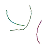
| ||||||||
|---|---|---|---|---|---|---|---|---|---|
| 1 | 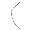
| ||||||||
| 2 | 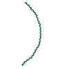
| ||||||||
| 3 | 
| ||||||||
| Unit cell |
|
-Components
-RNA chain , 6 types, 6 molecules IJKLMN
| #1: RNA chain | Mass: 135587.469 Da / Num. of mol.: 1 / Source method: isolated from a natural source / Source: (natural) BLUETONGUE VIRUS / Strain: STEROTYPE 1 (SOUTH AFRICA) |
|---|---|
| #2: RNA chain | Mass: 90815.789 Da / Num. of mol.: 1 / Source method: isolated from a natural source / Source: (natural) BLUETONGUE VIRUS / Strain: STEROTYPE 1 (SOUTH AFRICA) |
| #3: RNA chain | Mass: 87194.547 Da / Num. of mol.: 1 / Source method: isolated from a natural source / Source: (natural) BLUETONGUE VIRUS / Strain: STEROTYPE 1 (SOUTH AFRICA) |
| #4: RNA chain | Mass: 126095.031 Da / Num. of mol.: 1 / Source method: isolated from a natural source / Source: (natural) BLUETONGUE VIRUS / Strain: STEROTYPE 1 (SOUTH AFRICA) |
| #5: RNA chain | Mass: 84456.609 Da / Num. of mol.: 1 / Source method: isolated from a natural source / Source: (natural) BLUETONGUE VIRUS / Strain: STEROTYPE 1 (SOUTH AFRICA) |
| #6: RNA chain | Mass: 81088.797 Da / Num. of mol.: 1 / Source method: isolated from a natural source / Source: (natural) BLUETONGUE VIRUS / Strain: STEROTYPE 1 (SOUTH AFRICA) |
-Details
| Compound details | THIS ENTRY CORRESPONDS TO ONLY THE RNA MODEL WHICH WAS BUILT INTO THE BLUE TONGUE VIRUS STRUCTURE ...THIS ENTRY CORRESPOND |
|---|---|
| Sequence details | THIS SUBMISSION IS AN RNA MODEL THAT HAS BEEN RIGID BODY FITTED INTO A 10A DIFFERENCE FOURIER ...THIS SUBMISSION |
-Experimental details
-Experiment
| Experiment | Method: X-RAY DIFFRACTION / Number of used crystals: 1045 |
|---|
- Sample preparation
| Crystal | Description: OVER 1000 CRYSTALS EXAMINED. DATA WERE COLLECTED DURING 1995-1997. WAVELENGTHS BETWEEN 0.87 - 1.00. | ||||||||||||||||||||||||
|---|---|---|---|---|---|---|---|---|---|---|---|---|---|---|---|---|---|---|---|---|---|---|---|---|---|
| Crystal grow | pH: 8 Details: 15% AMMONIUM SULPHATE, 25% ETHYLENE GLYCOL, 0.1M TRIS/HCL BUFFER PH 8.0 | ||||||||||||||||||||||||
| Crystal grow | *PLUS pH: 8 / Method: vapor diffusion, sitting drop / Details: Diprose, J.M., (2001) EMBO J., 20, 7229. | ||||||||||||||||||||||||
| Components of the solutions | *PLUS
|
-Data collection
| Diffraction | Mean temperature: 281 K |
|---|---|
| Diffraction source | Source: SYNCHROTRON / Site: ESRF  / Beamline: ID2 / Wavelength: 1 / Beamline: ID2 / Wavelength: 1 |
| Detector | Type: MARRESEARCH / Detector: IMAGE PLATE |
| Radiation | Monochromator: DIAMOND / Protocol: SINGLE WAVELENGTH / Monochromatic (M) / Laue (L): M / Scattering type: x-ray |
| Radiation wavelength | Wavelength: 1 Å / Relative weight: 1 |
| Reflection | Resolution: 3.5→100 Å / Num. obs: 3524397 / % possible obs: 54 % / Redundancy: 6.5 % / Rmerge(I) obs: 0.229 / Net I/σ(I): 2.9 |
| Reflection shell | Resolution: 3.5→4 Å / Redundancy: 1.06 % / Rmerge(I) obs: 0.6 / Mean I/σ(I) obs: 0.72 / % possible all: 7.8 |
| Reflection | *PLUS Highest resolution: 10 Å / Lowest resolution: 105 Å / Num. obs: 251202 / % possible obs: 94.7 % / Num. measured all: 490727 / Rmerge(I) obs: 0.1 |
- Processing
| Software |
| ||||||||||||
|---|---|---|---|---|---|---|---|---|---|---|---|---|---|
| Refinement | Method to determine structure: MOLECULAR REPLACEMENT / Highest resolution: 10 Å Details: THE MODEL WAS BUILT INTO A 10A DIFFERENCE FOURIER ELECTRON DENSITY MAP CALCULATED BETWEEN NATIVE BLUETONGUE CORE DATA AND THE MODEL FOR THE CORE PARTICLE. CNS WAS USED TO RIGID BODY REFINE ...Details: THE MODEL WAS BUILT INTO A 10A DIFFERENCE FOURIER ELECTRON DENSITY MAP CALCULATED BETWEEN NATIVE BLUETONGUE CORE DATA AND THE MODEL FOR THE CORE PARTICLE. CNS WAS USED TO RIGID BODY REFINE THE MODEL AGAINST THE STRUCTURE FACTOR AMPLITUDES CALCULATED FROM THE DIFFERENCE MAP USING BASE PAIRS AS RIGID BODIES. | ||||||||||||
| Refinement step | Cycle: LAST / Highest resolution: 10 Å
|
