 Movie
Movie Controller


+ Open data

- Basic information
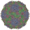
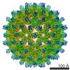
| Entry | Database: PDB / ID: 1al0 | ||||||
|---|---|---|---|---|---|---|---|
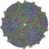
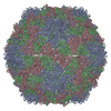
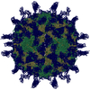
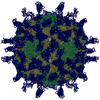
| Title | PROCAPSID OF BACTERIOPHAGE PHIX174 | ||||||
 Components Components |
| ||||||
 Keywords Keywords | VIRUS / COMPLEX (VIRUS CAPSID PROTEINS) / BACTERIOPHAGE / PROCAPSID / SCAFFOLDING PROTEIN / CHAPERONE / Icosahedral virus | ||||||
| Function / homology |  Function and homology information Function and homology informationviral scaffold assembly and maintenance / viral scaffold / symbiont-mediated perturbation of host process / viral procapsid maturation / Hydrolases; Acting on peptide bonds (peptidases) / T=1 icosahedral viral capsid / viral capsid / peptidase activity / host cell cytoplasm / symbiont entry into host cell ...viral scaffold assembly and maintenance / viral scaffold / symbiont-mediated perturbation of host process / viral procapsid maturation / Hydrolases; Acting on peptide bonds (peptidases) / T=1 icosahedral viral capsid / viral capsid / peptidase activity / host cell cytoplasm / symbiont entry into host cell / virion attachment to host cell / structural molecule activity / proteolysis Similarity search - Function | ||||||
| Biological species |  Enterobacteria phage phiX174 (virus) Enterobacteria phage phiX174 (virus) | ||||||
| Method |  X-RAY DIFFRACTION / SYNCHROTRON / MOLECULAR REPLACEMENT / Resolution: 3.5 Å X-RAY DIFFRACTION / SYNCHROTRON / MOLECULAR REPLACEMENT / Resolution: 3.5 Å | ||||||
 Authors Authors | Rossmann, M.G. / Dokland, T. | ||||||
 Citation Citation | Journal: Nature / Year: 1997 Title: Structure of a viral procapsid with molecular scaffolding. Authors: Dokland, T. / McKenna, R. / Ilag, L.L. / Bowman, B.R. / Incardona, N.L. / Fane, B.A. / Rossmann, M.G. #1: Journal: Structure / Year: 1995Title: DNA Packaging Intermediates of Bacteriophage Phi X174 Authors: Ilag, L.L. / Olson, N.H. / Dokland, T. / Music, C.L. / Cheng, R.H. / Bowen, Z. / McKenna, R. / Rossmann, M.G. / Baker, T.S. / Incardona, N.L. #2: Journal: J.Mol.Biol. / Year: 1994Title: Analysis of the Single-Stranded DNA Bacteriophage Phi X174, Refined at a Resolution of 3.0 A Authors: McKenna, R. / Ilag, L.L. / Rossmann, M.G. #3: Journal: Nature / Year: 1992Title: Atomic Structure of Single-Stranded DNA Bacteriophage Phi X174 and its Functional Implications Authors: McKenna, R. / Xia, D. / Willingmann, P. / Ilag, L.L. / Krishnaswamy, S. / Rossmann, M.G. / Olson, N.H. / Baker, T.S. / Incardona, N.L. #4: Journal: The Bacteriophages (The Viruses) / Year: 1988Title: Biology of the Bacteriophage PhiX174 Authors: Hayashi, M. / Aoyama, A. / Delwood, L. / Richardson, D.L. / Hayashi, M.N. #5: Journal: Nature / Year: 1977Title: Nucleotide Sequence of Bacteriophage Phi X174 DNA Authors: Sanger, F. / Air, G.M. / Barrell, B.G. / Brown, N.L. / Coulson, A.R. / Fiddes, J.C. / Hutchison, C.A. / Slocombe, P.M. / Smith, M. | ||||||
| History |
| ||||||
| Remark 285 | THE ENTRY PRESENTED HERE DOES NOT CONTAIN THE COMPLETE CRYSTAL ASYMMETRIC UNIT. IN ADDITION, THE ...THE ENTRY PRESENTED HERE DOES NOT CONTAIN THE COMPLETE CRYSTAL ASYMMETRIC UNIT. IN ADDITION, THE COORDINATES ARE NOT PRESENTED IN THE STANDARD CRYSTAL FRAME. IN ORDER TO GENERATE THE FULL CRYSTAL AU, APPLY THE FOLLOWING TRANSFORMATION MATRIX OR MATRICES AND SELECTED BIOMT RECORDS TO THE COORDINATES, AS SHOWN BELOW. X0 1 1.000000 0.000000 0.000000 188.08200 X0 2 0.000000 1.000000 0.000000 188.08200 X0 3 0.000000 0.000000 1.000000 188.08200 X1 1 0.834253 0.463850 -0.298103 -4.02480 X1 2 -0.298103 0.834253 0.463850 -4.02480 X1 3 0.463850 -0.298103 0.834253 -4.02480 CRYSTAL AU = (X0) * (BIOMT 1-20) * CHAINS 1,2,3,4,F,G,B + (X1) * (BIOMT 1-20) * CHAINS 1,2,3,4,F,G,B |
- Structure visualization
| Structure viewer | Molecule: MolmilJmol/JSmol |
|---|
- Downloads & links
-Download
| PDBx/mmCIF format | 1al0.cif.gz | 219.1 KB | Display | PDBx/mmCIF format |
|---|---|---|---|---|
| PDB format | pdb1al0.ent.gz | 169.2 KB | Display | PDB format |
| PDBx/mmJSON format | 1al0.json.gz | Tree view | PDBx/mmJSON format | |
| Others |  Other downloads Other downloads |
-Validation report
| Arichive directory | https://data.pdbj.org/pub/pdb/validation_reports/al/1al0ftp://data.pdbj.org/pub/pdb/validation_reports/al/1al0 | HTTPS FTP |
|---|
-Related structure data
| Related structure data |  1phxS S: Starting model for refinement |
|---|---|
| Similar structure data |


-Links
 PDBj
PDBj


- Assembly
| Deposited unit | 
| ||||||||
|---|---|---|---|---|---|---|---|---|---|
| 1 | x 60
| ||||||||
| 2 |
| ||||||||
| 3 | x 5
| ||||||||
| 4 | x 6
| ||||||||
| 5 | 
| ||||||||
| 6 | x 20 x 20 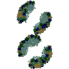
| ||||||||
| Unit cell |
| ||||||||
| Symmetry | Point symmetry: (Hermann–Mauguin notation: 532 / Schoenflies symbol: I (icosahedral)) |
-Components
| #1: Protein | Mass: 16953.316 Da / Num. of mol.: 4 / Source method: isolated from a natural source / Source: (natural) Enterobacteria phage phiX174 (virus) / Genus: Microvirus / Species: Enterobacteria phage phiX174 sensu lato / Strain: C / References: UniProt: P69486#2: Protein | | Mass: 48423.375 Da / Num. of mol.: 1 / Source method: isolated from a natural source / Source: (natural) Enterobacteria phage phiX174 (virus) / Genus: Microvirus / Species: Enterobacteria phage phiX174 sensu lato / Strain: C / References: UniProt: P03641#3: Protein | | Mass: 19061.652 Da / Num. of mol.: 1 / Source method: isolated from a natural source / Source: (natural) Enterobacteria phage phiX174 (virus) / Genus: Microvirus / Species: Enterobacteria phage phiX174 sensu lato / Strain: C / References: UniProt: P03643#4: Protein | | Mass: 13863.118 Da / Num. of mol.: 1 / Source method: isolated from a natural source / Source: (natural) Enterobacteria phage phiX174 (virus) / Genus: Microvirus / Species: Enterobacteria phage phiX174 sensu lato / Strain: C / References: UniProt: P03633 |
|---|
-Experimental details
-Experiment
| Experiment | Method: X-RAY DIFFRACTION / Number of used crystals: 22 |
|---|
- Sample preparation
| Crystal grow | Method: vapor diffusion / pH: 6 Details: PROCAPSIDS WERE CRYSTALLIZED BY VAPOUR DIFFUSION FROM 43-37% (OF SATURATION) AMMONIUM SULFATE, 100MM MES PH6.0, vapor diffusion | ||||||||||||||||||||||||||||||||||||||||||||||||
|---|---|---|---|---|---|---|---|---|---|---|---|---|---|---|---|---|---|---|---|---|---|---|---|---|---|---|---|---|---|---|---|---|---|---|---|---|---|---|---|---|---|---|---|---|---|---|---|---|---|
| Crystal grow | *PLUS pH: 7.5 / Method: vapor diffusion, hanging drop | ||||||||||||||||||||||||||||||||||||||||||||||||
| Components of the solutions | *PLUS
|
-Data collection
| Diffraction | Mean temperature: 277 K |
|---|---|
| Diffraction source | Source: SYNCHROTRON / Site: CHESS  / Beamline: F1 / Wavelength: 0.918 / Beamline: F1 / Wavelength: 0.918 |
| Detector | Type: FUJI / Detector: IMAGE PLATE / Date: Jan 1, 1996 |
| Radiation | Monochromatic (M) / Laue (L): M / Scattering type: x-ray |
| Radiation wavelength | Wavelength: 0.918 Å / Relative weight: 1 |
| Reflection | Resolution: 3.5→45 Å / Num. obs: 527445 / % possible obs: 55.3 % / Observed criterion σ(I): -3 / Redundancy: 2 % / Rmerge(I) obs: 0.247 / Rsym value: 0.247 |
| Reflection | *PLUS Num. all: 1072459 / Num. measured all: 1646040 |
- Processing
| Software |
| ||||||||||||||||||||||||
|---|---|---|---|---|---|---|---|---|---|---|---|---|---|---|---|---|---|---|---|---|---|---|---|---|---|
| Refinement | Method to determine structure: MOLECULAR REPLACEMENT Starting model: PDB ENTRY 1PHX Resolution: 3.5→8 Å / Data cutoff high absF: 100000 / Data cutoff low absF: 0.1 / σ(F): 2 /
| ||||||||||||||||||||||||
| Refinement step | Cycle: LAST / Resolution: 3.5→8 Å
| ||||||||||||||||||||||||
| Refine LS restraints NCS | NCS model details: CONSTRAINTS | ||||||||||||||||||||||||
| LS refinement shell | Resolution: 3.5→3.64 Å / Total num. of bins used: 8 /
| ||||||||||||||||||||||||
| Xplor file |
|
