 Movie
Movie Controller

[English] 日本語
 Yorodumi
- PDB-1agb: ANTAGONIST HIV-1 GAG PEPTIDES INDUCE STRUCTURAL CHANGES IN HLA B8... -
+ Open data

- Basic information
| Entry | Database: PDB / ID: 1agb | ||||||
|---|---|---|---|---|---|---|---|
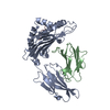
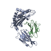
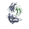
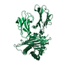
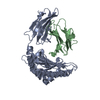
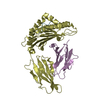
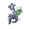
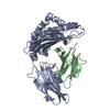
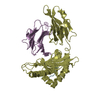
| Title | ANTAGONIST HIV-1 GAG PEPTIDES INDUCE STRUCTURAL CHANGES IN HLA B8-HIV-1 GAG PEPTIDE (GGRKKYKL-3R MUTATION) | ||||||
 Components Components |
| ||||||
 Keywords Keywords | HISTOCOMPATIBILITY COMPLEX / HLA B8 / HIV / MHC CLASS I | ||||||
| Function / homology |  Function and homology information Function and homology informationregulation of interleukin-12 production / regulation of dendritic cell differentiation / regulation of T cell anergy / regulation of interleukin-6 production / TAP binding / protection from natural killer cell mediated cytotoxicity / detection of bacterium / positive regulation of ferrous iron binding / positive regulation of transferrin receptor binding / secretory granule membrane ...regulation of interleukin-12 production / regulation of dendritic cell differentiation / regulation of T cell anergy / regulation of interleukin-6 production / TAP binding / protection from natural killer cell mediated cytotoxicity / detection of bacterium / positive regulation of ferrous iron binding / positive regulation of transferrin receptor binding / secretory granule membrane / negative regulation of receptor binding / DAP12 interactions / antigen processing and presentation of endogenous peptide antigen via MHC class I via ER pathway, TAP-independent / positive regulation of receptor binding / antigen processing and presentation of endogenous peptide antigen via MHC class Ib / early endosome lumen / Nef mediated downregulation of MHC class I complex cell surface expression / cellular response to iron ion / Endosomal/Vacuolar pathway / lumenal side of endoplasmic reticulum membrane / Antigen Presentation: Folding, assembly and peptide loading of class I MHC / cellular response to iron(III) ion / antigen processing and presentation of exogenous protein antigen via MHC class Ib, TAP-dependent / negative regulation of forebrain neuron differentiation / regulation of erythrocyte differentiation / peptide antigen assembly with MHC class I protein complex / ER to Golgi transport vesicle membrane / regulation of iron ion transport / response to molecule of bacterial origin / MHC class I peptide loading complex / HFE-transferrin receptor complex / T cell mediated cytotoxicity / positive regulation of T cell cytokine production / antigen processing and presentation of endogenous peptide antigen via MHC class I / sensory perception of smell / MHC class I protein complex / defense response / negative regulation of neurogenesis / multicellular organismal-level iron ion homeostasis / positive regulation of receptor-mediated endocytosis / peptide antigen assembly with MHC class II protein complex / MHC class II protein complex / cellular response to nicotine / positive regulation of T cell mediated cytotoxicity / specific granule lumen / phagocytic vesicle membrane / recycling endosome membrane / peptide antigen binding / positive regulation of cellular senescence / antigen processing and presentation of exogenous peptide antigen via MHC class II / negative regulation of epithelial cell proliferation / Immunoregulatory interactions between a Lymphoid and a non-Lymphoid cell / Interferon gamma signaling / positive regulation of immune response / positive regulation of T cell activation / Modulation by Mtb of host immune system / Interferon alpha/beta signaling / negative regulation of neuron projection development / positive regulation of protein binding / tertiary granule lumen / DAP12 signaling / MHC class II protein complex binding / late endosome membrane / protein-folding chaperone binding / iron ion transport / ER-Phagosome pathway / early endosome membrane / T cell differentiation in thymus / protein refolding / protein homotetramerization / intracellular iron ion homeostasis / adaptive immune response / amyloid fibril formation / learning or memory / immune response / Amyloid fiber formation / endoplasmic reticulum lumen / lysosomal membrane / external side of plasma membrane / Golgi membrane / innate immune response / signaling receptor binding / focal adhesion / Neutrophil degranulation / SARS-CoV-2 activates/modulates innate and adaptive immune responses / structural molecule activity / Golgi apparatus / cell surface / endoplasmic reticulum / protein homodimerization activity / extracellular space / extracellular exosome / extracellular region / identical protein binding / membrane / plasma membrane / cytosol Similarity search - Function | ||||||
| Biological species |  Homo sapiens (human) Homo sapiens (human)  Human immunodeficiency virus 1 Human immunodeficiency virus 1 | ||||||
| Method |  X-RAY DIFFRACTION / SYNCHROTRON / MOLECULAR REPLACEMENT / Resolution: 2.2 Å X-RAY DIFFRACTION / SYNCHROTRON / MOLECULAR REPLACEMENT / Resolution: 2.2 Å | ||||||
 Authors Authors | Reid, S.W. / Mcadam, S. / Smith, K.J. / Klenerman, P. / O'Callaghan, C.A. / Harlos, K. / Jakobsen, B.K. / Mcmichael, A.J. / Bell, J. / Stuart, D.I. / Jones, E.Y. | ||||||
 Citation Citation | Journal: J.Exp.Med. / Year: 1996 Title: Antagonist HIV-1 Gag peptides induce structural changes in HLA B8. Authors: Reid, S.W. / McAdam, S. / Smith, K.J. / Klenerman, P. / O'Callaghan, C.A. / Harlos, K. / Jakobsen, B.K. / McMichael, A.J. / Bell, J.I. / Stuart, D.I. / Jones, E.Y. | ||||||
| History |
|
- Structure visualization
| Structure viewer | Molecule: MolmilJmol/JSmol |
|---|
- Downloads & links
-Download
| PDBx/mmCIF format | 1agb.cif.gz | 96.6 KB | Display | PDBx/mmCIF format |
|---|---|---|---|---|
| PDB format | pdb1agb.ent.gz | 73.8 KB | Display | PDB format |
| PDBx/mmJSON format | 1agb.json.gz | Tree view | PDBx/mmJSON format | |
| Others |  Other downloads Other downloads |
-Validation report
| Summary document | 1agb_validation.pdf.gz | 373.9 KB | Display | wwPDB validaton report |
|---|---|---|---|---|
| Full document | 1agb_full_validation.pdf.gz | 376.7 KB | Display | |
| Data in XML | 1agb_validation.xml.gz | 8.7 KB | Display | |
| Data in CIF | 1agb_validation.cif.gz | 14.8 KB | Display | |
| Arichive directory | https://data.pdbj.org/pub/pdb/validation_reports/ag/1agbftp://data.pdbj.org/pub/pdb/validation_reports/ag/1agb | HTTPS FTP |
-Related structure data


-Links
 PDBj
PDBj


- Assembly
| Deposited unit | 
| ||||||||
|---|---|---|---|---|---|---|---|---|---|
| 1 |
| ||||||||
| Unit cell |
|
-Components
| #1: Protein | Mass: 31927.977 Da / Num. of mol.: 1 / Fragment: EXTRACELLULAR Source method: isolated from a genetically manipulated source Source: (gene. exp.) Homo sapiens (human) / Cell line: XA90 / Gene: GAG / Plasmid: PGMT7 / Cell line (production host): BL21(DE3)PLYSS / Gene (production host): GAG / Production host:  |
|---|---|
| #2: Protein | Mass: 11748.160 Da / Num. of mol.: 1 / Fragment: EXTRACELLULAR Source method: isolated from a genetically manipulated source Source: (gene. exp.) Homo sapiens (human) / Cell line: XA90 / Gene: GAG / Plasmid: PHN1 / Cell line (production host): XA90 / Gene (production host): GAG / Production host: |
| #3: Protein/peptide | Mass: 953.184 Da / Num. of mol.: 1 / Fragment: EXTRACELLULAR Source method: isolated from a genetically manipulated source Source: (gene. exp.) Human immunodeficiency virus 1 / Genus: Lentivirus |
| #4: Water | ChemComp-HOH /  Mass: 18.015 Da / Num. of mol.: 306 / Source method: isolated from a natural source / Formula: H2O Mass: 18.015 Da / Num. of mol.: 306 / Source method: isolated from a natural source / Formula: H2O |
-Experimental details
-Experiment
| Experiment | Method: X-RAY DIFFRACTION / Number of used crystals: 1 |
|---|
- Sample preparation
| Crystal | Density Matthews: 2.6 Å3/Da / Density % sol: 52.7 % | ||||||||||||||||||||||||||||||
|---|---|---|---|---|---|---|---|---|---|---|---|---|---|---|---|---|---|---|---|---|---|---|---|---|---|---|---|---|---|---|---|
| Crystal grow | pH: 6.5 Details: 30% PEG 4000 0.1M SODIUM CITRATE, PH 6.5 0.2M AMMONIUM ACETATE | ||||||||||||||||||||||||||||||
| Crystal grow | *PLUS Temperature: 21 ℃ / pH: 8 / Method: vapor diffusion, sitting drop / Details: Reid, S.W., (1996) Febs Lett., 383, 119. | ||||||||||||||||||||||||||||||
| Components of the solutions | *PLUS
|
-Data collection
| Diffraction | Mean temperature: 187 K |
|---|---|
| Diffraction source | Source: SYNCHROTRON / Site: SRS  / Beamline: PX9.6 / Wavelength: 0.97 / Beamline: PX9.6 / Wavelength: 0.97 |
| Detector | Type: MARRESEARCH / Detector: IMAGE PLATE / Date: Sep 2, 1996 |
| Radiation | Monochromatic (M) / Laue (L): M / Scattering type: x-ray |
| Radiation wavelength | Wavelength: 0.97 Å / Relative weight: 1 |
| Reflection | Resolution: 2.2→14 Å / Num. obs: 27424 / % possible obs: 97.7 % / Observed criterion σ(I): 0 / Redundancy: 3.2 % / Rmerge(I) obs: 0.089 / Net I/σ(I): 8.5 |
| Reflection shell | Resolution: 2.2→2.3 Å / Rmerge(I) obs: 0.232 / % possible all: 91.5 |
| Reflection | *PLUS Num. measured all: 88881 |
- Processing
| Software |
| |||||||||||||||||||||||||||||||||||||||||||||||||||||||||||||||
|---|---|---|---|---|---|---|---|---|---|---|---|---|---|---|---|---|---|---|---|---|---|---|---|---|---|---|---|---|---|---|---|---|---|---|---|---|---|---|---|---|---|---|---|---|---|---|---|---|---|---|---|---|---|---|---|---|---|---|---|---|---|---|---|---|
| Refinement | Method to determine structure: MOLECULAR REPLACEMENT Starting model: HLA B27 Resolution: 2.2→14 Å / σ(F): 0
| |||||||||||||||||||||||||||||||||||||||||||||||||||||||||||||||
| Displacement parameters | Biso mean: 21.1 Å2 | |||||||||||||||||||||||||||||||||||||||||||||||||||||||||||||||
| Refinement step | Cycle: LAST / Resolution: 2.2→14 Å
| |||||||||||||||||||||||||||||||||||||||||||||||||||||||||||||||
| Refine LS restraints |
| |||||||||||||||||||||||||||||||||||||||||||||||||||||||||||||||
| Software | *PLUS Name: PROLSQ / Classification: refinement | |||||||||||||||||||||||||||||||||||||||||||||||||||||||||||||||
| Refinement | *PLUS Rfactor all: 0.195 | |||||||||||||||||||||||||||||||||||||||||||||||||||||||||||||||
| Solvent computation | *PLUS | |||||||||||||||||||||||||||||||||||||||||||||||||||||||||||||||
| Displacement parameters | *PLUS |
