Flagellar hook protein FlgE superfamily / Flagellar hook protein FlgE / Flagellar basal body protein FlaE D2 domain / Flagellar hook-basal body protein, FlgE/F/G / Flagellar hook-basal body protein, FlgE/F/G-like / : / Flagellar hook protein FlgE/F/G D1 domain / Flagellar basal body rod protein, conserved site / Flagella basal body rod proteins signature. / Flagellar basal body rod protein, N-terminal ...Flagellar hook protein FlgE superfamily / Flagellar hook protein FlgE / Flagellar basal body protein FlaE D2 domain / Flagellar hook-basal body protein, FlgE/F/G / Flagellar hook-basal body protein, FlgE/F/G-like / : / Flagellar hook protein FlgE/F/G D1 domain / Flagellar basal body rod protein, conserved site / Flagella basal body rod proteins signature. / Flagellar basal body rod protein, N-terminal / Flagellar basal-body/hook protein, C-terminal domain / Flagella basal body rod protein / Flagellar basal body rod FlgEFG protein C-terminal Similarity search - Domain/homology
Japan Agency for Medical Research and Development (AMED)
JP18am0101076
Japan
Japan Society for the Promotion of Science
17K17085
Japan
Japan Society for the Promotion of Science
19K10083
Japan
Japan Society for the Promotion of Science
17K07318
Japan
Citation
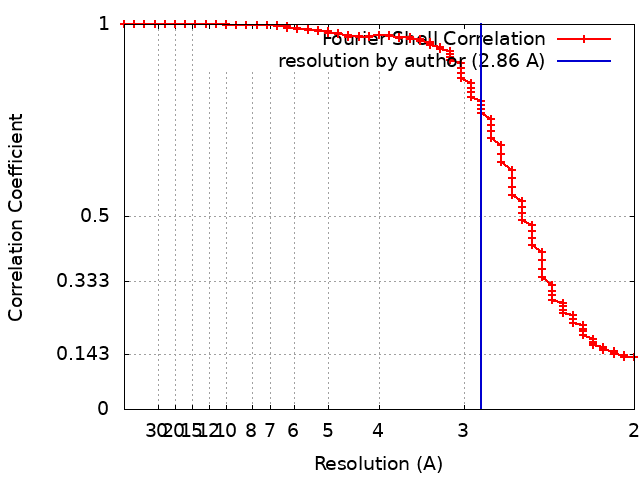
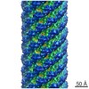
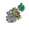
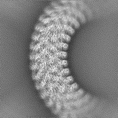
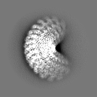
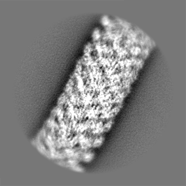
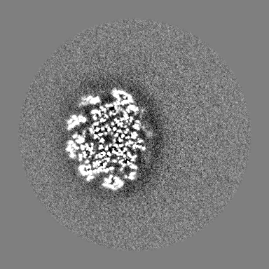
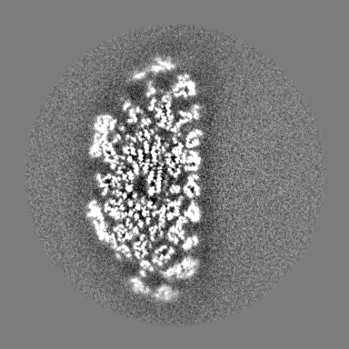
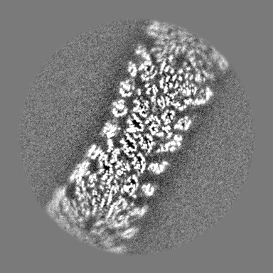
Journal: Nat Struct Mol Biol / Year: 2019 Title: Torque transmission mechanism of the curved bacterial flagellar hook revealed by cryo-EM. Authors: Satoshi Shibata / Hideyuki Matsunami / Shin-Ichi Aizawa / Matthias Wolf / Abstract: Bacterial locomotion by rotating flagella is achieved through the hook, which transmits torque from the motor to the filament. The hook is a tubular structure composed of a single type of protein, ...Bacterial locomotion by rotating flagella is achieved through the hook, which transmits torque from the motor to the filament. The hook is a tubular structure composed of a single type of protein, yet it adopts a curved shape. To perform its function, it must be simultaneously flexible and torsionally rigid. The molecular mechanism by which chemically identical subunits form such a dynamic structure is unknown. Here, we show the complete structure of the hook from Salmonella enterica in its supercoiled 'curved' state, at 2.9 Å resolution. Subunits in the curved hook are grouped into 11 distinctive conformations, each shared along 11 protofilaments. The domains of the elongated hook subunit behave as rigid bodies connected by two hinge regions. The reconstituted model demonstrates how identical subunits can dynamically change conformation by physical interactions while bending. These multiple subunit states contradict the two-state model, which is a key feature of flagellar polymorphism.
History
Deposition
May 19, 2019
-
Header (metadata) release
Oct 2, 2019
-
Map release
Oct 2, 2019
-
Update
Mar 27, 2024
-
Current status
Mar 27, 2024
Processing site: PDBj / Status: Released
-
Structure visualization
Movie
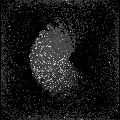
Surface view with section colored by density value
Cryogen name: ETHANE / Chamber humidity: 100 % / Chamber temperature: 289 K / Instrument: FEI VITROBOT MARK IV / Details: 3 second blot, 4.0uL.
-
Electron microscopy
Microscope
FEI TITAN KRIOS
Temperature
Min: 77.0 K / Max: 100.0 K
Details
nanoprobe, parallel beam illumination
Image recording
Film or detector model: FEI FALCON III (4k x 4k) / Detector mode: INTEGRATING / Digitization - Dimensions - Width: 4000 pixel / Digitization - Dimensions - Height: 4000 pixel / Number grids imaged: 1 / Number real images: 2655 / Average exposure time: 2.0 sec. / Average electron dose: 89.0 e/Å2 Details: frame alignment and dose weighting using motioncor2
Electron beam
Acceleration voltage: 300 kV / Electron source: FIELD EMISSION GUN
In the structure databanks used in Yorodumi, some data are registered as the other names, "COVID-19 virus" and "2019-nCoV". Here are the details of the virus and the list of structure data.
Jan 31, 2019. EMDB accession codes are about to change! (news from PDBe EMDB page)
EMDB accession codes are about to change! (news from PDBe EMDB page)
The allocation of 4 digits for EMDB accession codes will soon come to an end. Whilst these codes will remain in use, new EMDB accession codes will include an additional digit and will expand incrementally as the available range of codes is exhausted. The current 4-digit format prefixed with “EMD-” (i.e. EMD-XXXX) will advance to a 5-digit format (i.e. EMD-XXXXX), and so on. It is currently estimated that the 4-digit codes will be depleted around Spring 2019, at which point the 5-digit format will come into force.
The EM Navigator/Yorodumi systems omit the EMD- prefix.
Related info.:Q: What is EMD? / ID/Accession-code notation in Yorodumi/EM Navigator
Yorodumi is a browser for structure data from EMDB, PDB, SASBDB, etc.
This page is also the successor to EM Navigator detail page, and also detail information page/front-end page for Omokage search.
The word "yorodu" (or yorozu) is an old Japanese word meaning "ten thousand". "mi" (miru) is to see.
Related info.:EMDB / PDB / SASBDB / Comparison of 3 databanks / Yorodumi Search / Aug 31, 2016. New EM Navigator & Yorodumi / Yorodumi Papers / Jmol/JSmol / Function and homology information / Changes in new EM Navigator and Yorodumi
 Movie
Movie Controller


 Open data
 Basic information
 Map data
 Sample
 Keywords
 Function and homology information
 Salmonella enterica subsp. enterica serovar Typhimurium str. LT2 (bacteria) /
  Authors
 Japan, 4 items
  Citation
 Structure visualization
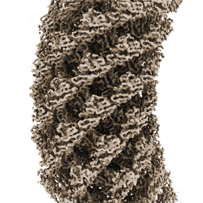
 Downloads & links
 emd_9909.png
 http://ftp.pdbj.org/pub/emdb/structures/EMD-9909


 Z (Sec.)
 Y (Row.)
 X (Col.)


 Sample components
 Processing
 Electron microscopy
 FIELD EMISSION GUN