Movie
Movie Controller

[English] 日本語
 Yorodumi
- EMDB-9374: Single particle reconstruction of DARPin and its bound GFP on a s... -
+ Open data

- Basic information
| Entry | Database: EMDB / ID: EMD-9374 | |||||||||||||||||||||
|---|---|---|---|---|---|---|---|---|---|---|---|---|---|---|---|---|---|---|---|---|---|---|
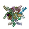
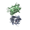
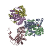
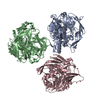
| Title | Single particle reconstruction of DARPin and its bound GFP on a symmetric scaffold | |||||||||||||||||||||
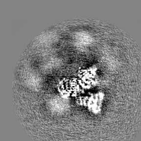
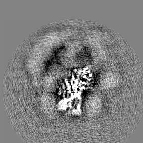
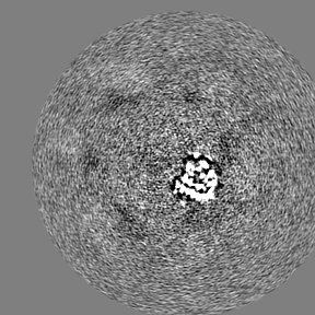
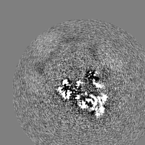
 Map data Map data | Refinement of DARPin, GFP, and two adjacent trimers | |||||||||||||||||||||
 Sample Sample |
| |||||||||||||||||||||
 Keywords Keywords | protein engineering / symmetric scaffold / small protein cryo-EM / display platform / BIOSYNTHETIC PROTEIN | |||||||||||||||||||||
| Function / homology | 5-carboxymethyl-2-hydroxymuconate isomerase / 5-carboxymethyl-2-hydroxymuconate isomerase / 5-carboxymethyl-2-hydroxymuconate delta-isomerase activity / Tautomerase/MIF superfamily / 5-carboxymethyl-2-hydroxymuconate isomerase Function and homology information Function and homology information | |||||||||||||||||||||
| Biological species |   Aequorea victoria (jellyfish) / Aequorea victoria (jellyfish) /   Pyrococcus horikoshii OT3 (archaea) / Pyrococcus horikoshii OT3 (archaea) /  Pseudomonas aeruginosa PAO1 (bacteria) / Pseudomonas aeruginosa (strain ATCC 15692 / DSM 22644 / CIP 104116 / JCM 14847 / LMG 12228 / 1C / PRS 101 / PAO1) (bacteria) / Pyrococcus horikoshii (strain ATCC 700860 / DSM 12428 / JCM 9974 / NBRC 100139 / OT-3) (archaea) Pseudomonas aeruginosa PAO1 (bacteria) / Pseudomonas aeruginosa (strain ATCC 15692 / DSM 22644 / CIP 104116 / JCM 14847 / LMG 12228 / 1C / PRS 101 / PAO1) (bacteria) / Pyrococcus horikoshii (strain ATCC 700860 / DSM 12428 / JCM 9974 / NBRC 100139 / OT-3) (archaea) | |||||||||||||||||||||
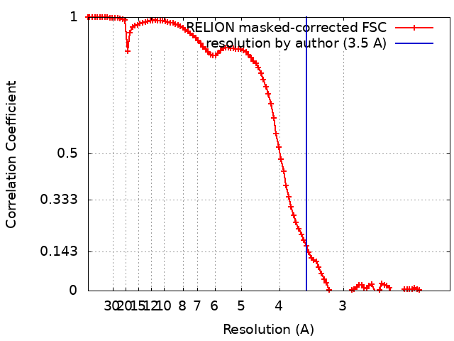
| Method | single particle reconstruction / cryo EM / Resolution: 3.5 Å | |||||||||||||||||||||
 Authors Authors | Liu Y / Huynh D | |||||||||||||||||||||
| Funding support |  United States, 6 items United States, 6 items
| |||||||||||||||||||||
 Citation Citation | Journal: Nat Commun / Year: 2019 Title: A 3.8 Å resolution cryo-EM structure of a small protein bound to an imaging scaffold. Authors: Yuxi Liu / Duc T Huynh / Todd O Yeates / Abstract: Proteins smaller than about 50 kDa are currently too small to be imaged at high resolution by cryo-electron microscopy (cryo-EM), leaving most protein molecules in the cell beyond the reach of this ...Proteins smaller than about 50 kDa are currently too small to be imaged at high resolution by cryo-electron microscopy (cryo-EM), leaving most protein molecules in the cell beyond the reach of this powerful structural technique. Here we use a designed protein scaffold to bind and symmetrically display 12 copies of a small 26 kDa protein, green fluorescent protein (GFP). We show that the bound cargo protein is held rigidly enough to visualize it at a resolution of 3.8 Å by cryo-EM, where specific structural features of the protein are visible. The designed scaffold is modular and can be modified through modest changes in its amino acid sequence to bind and display diverse proteins for imaging, thus providing a general method to break through the lower size limitation in cryo-EM. | |||||||||||||||||||||
| History |
|
- Structure visualization
| Movie |
Movie viewer |
|---|---|
| Structure viewer | EM map: SurfViewMolmilJmol/JSmol |
| Supplemental images |

- Downloads & links
-EMDB archive
| Map data | emd_9374.map.gz | 85.5 MB | EMDB map data format | |
|---|---|---|---|---|
| Header (meta data) | emd-9374-v30.xmlemd-9374.xml | 29.1 KB 29.1 KB | Display Display | EMDB header |
| FSC (resolution estimation) | emd_9374_fsc.xml | 10.3 KB | Display | FSC data file |
| Images | 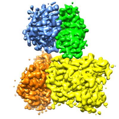 emd_9374.png emd_9374.png | 153.8 KB | ||
| Masks | emd_9374_msk_1.map | 91.1 MB | Mask map | |
| Filedesc metadata | emd-9374.cif.gz | 8 KB | ||
| Others | emd_9374_half_map_1.map.gzemd_9374_half_map_2.map.gz | 59.1 MB 59.1 MB | ||
| Archive directory |  http://ftp.pdbj.org/pub/emdb/structures/EMD-9374ftp://ftp.pdbj.org/pub/emdb/structures/EMD-9374 http://ftp.pdbj.org/pub/emdb/structures/EMD-9374ftp://ftp.pdbj.org/pub/emdb/structures/EMD-9374 | HTTPS FTP |
-Related structure data
| Related structure data |  6nhvMC 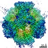 9373C  6nhtC C: citing same article ( M: atomic model generated by this map |
|---|---|
| Similar structure data |


-Links
| EMDB pages | EMDB (EBI/PDBe) / EMDataResource |
|---|
-Map
| File | Download / File: emd_9374.map.gz / Format: CCP4 / Size: 91.1 MB / Type: IMAGE STORED AS FLOATING POINT NUMBER (4 BYTES) | ||||||||||||||||||||||||||||||||||||||||||||||||||||||||||||||||||||
|---|---|---|---|---|---|---|---|---|---|---|---|---|---|---|---|---|---|---|---|---|---|---|---|---|---|---|---|---|---|---|---|---|---|---|---|---|---|---|---|---|---|---|---|---|---|---|---|---|---|---|---|---|---|---|---|---|---|---|---|---|---|---|---|---|---|---|---|---|---|
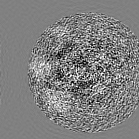
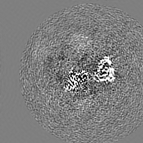
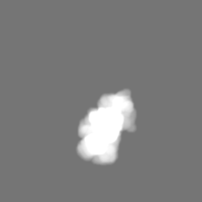
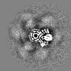
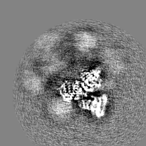
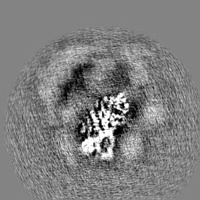
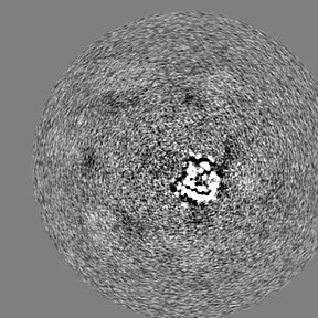
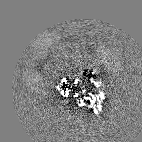
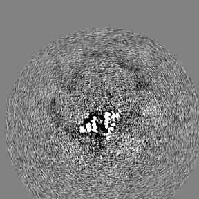
| Annotation | Refinement of DARPin, GFP, and two adjacent trimers | ||||||||||||||||||||||||||||||||||||||||||||||||||||||||||||||||||||
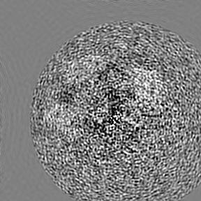
| Projections & slices | Image control
Images are generated by Spider. | ||||||||||||||||||||||||||||||||||||||||||||||||||||||||||||||||||||
| Voxel size | X=Y=Z: 1.06 Å | ||||||||||||||||||||||||||||||||||||||||||||||||||||||||||||||||||||
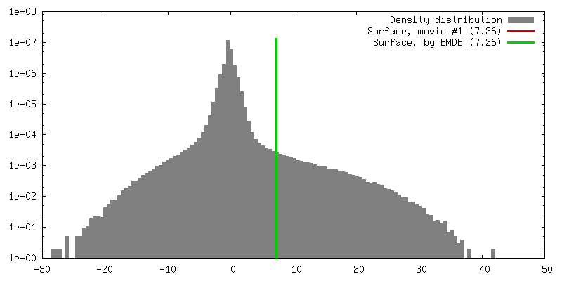
| Density |
| ||||||||||||||||||||||||||||||||||||||||||||||||||||||||||||||||||||
| Symmetry | Space group: 1 | ||||||||||||||||||||||||||||||||||||||||||||||||||||||||||||||||||||
| Details | EMDB XML:
CCP4 map header:
| ||||||||||||||||||||||||||||||||||||||||||||||||||||||||||||||||||||
 X (Sec.)
 Y (Row.)
 Z (Col.)


-Supplemental data
-Mask #1
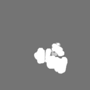
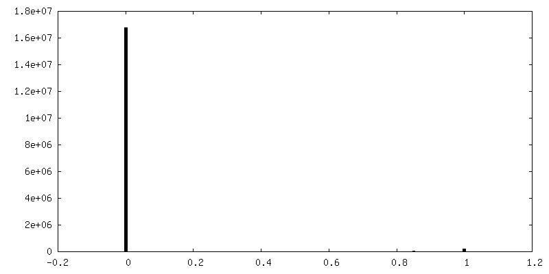
| File | emd_9374_msk_1.map | ||||||||||||
|---|---|---|---|---|---|---|---|---|---|---|---|---|---|
| Projections & Slices |
| ||||||||||||
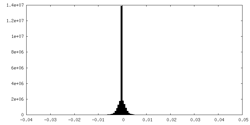
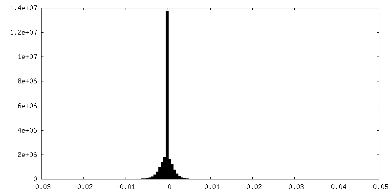
| Density Histograms |


-Half map: Half body 1
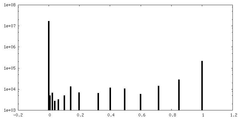
| File | emd_9374_half_map_1.map | ||||||||||||
|---|---|---|---|---|---|---|---|---|---|---|---|---|---|
| Annotation | Half body 1 | ||||||||||||
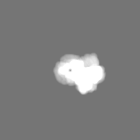
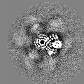
| Projections & Slices |
| ||||||||||||
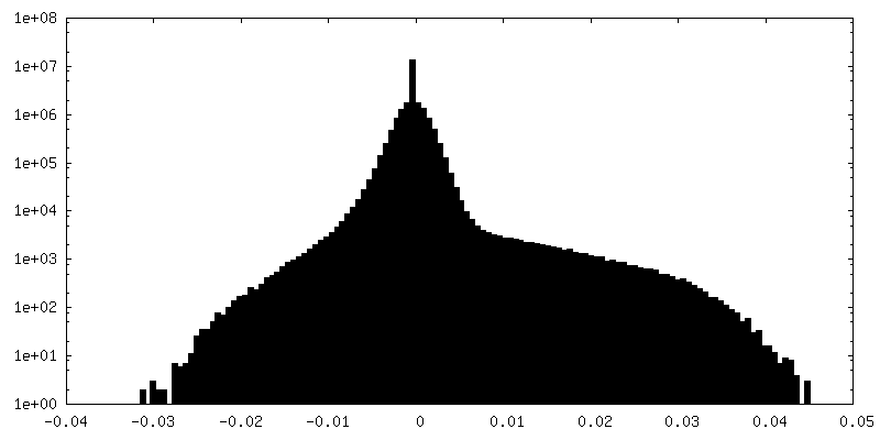
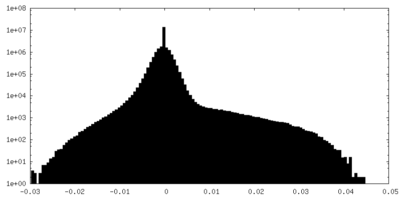
| Density Histograms |

-Half map: Half body 2
| File | emd_9374_half_map_2.map | ||||||||||||
|---|---|---|---|---|---|---|---|---|---|---|---|---|---|
| Annotation | Half body 2 | ||||||||||||
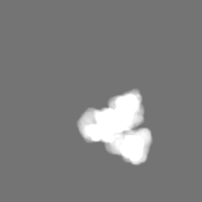
| Projections & Slices |
| ||||||||||||
| Density Histograms |
- Sample components
-Entire : Subunit A with DARPin + Subunit B + superfolder GFP
| Entire | Name: Subunit A with DARPin + Subunit B + superfolder GFP |
|---|---|
| Components |
|
-Supramolecule #1: Subunit A with DARPin + Subunit B + superfolder GFP
| Supramolecule | Name: Subunit A with DARPin + Subunit B + superfolder GFP / type: complex / ID: 1 / Parent: 0 / Macromolecule list: all |
|---|
-Supramolecule #2: superfolder GFP
| Supramolecule | Name: superfolder GFP / type: complex / ID: 2 / Parent: 1 / Macromolecule list: #1 |
|---|---|
| Source (natural) | Organism: Aequorea victoria (jellyfish) |
-Supramolecule #3: Subunit A with DARPin
| Supramolecule | Name: Subunit A with DARPin / type: complex / ID: 3 / Parent: 1 / Macromolecule list: #3 |
|---|---|
| Source (natural) | Organism: Pyrococcus horikoshii OT3 (archaea) |
-Supramolecule #4: Subunit B
| Supramolecule | Name: Subunit B / type: complex / ID: 4 / Parent: 1 / Macromolecule list: #2 |
|---|---|
| Source (natural) | Organism: Pseudomonas aeruginosa PAO1 (bacteria) |
-Macromolecule #1: superfolder GFP
| Macromolecule | Name: superfolder GFP / type: protein_or_peptide / ID: 1 / Number of copies: 1 / Enantiomer: LEVO |
|---|---|
| Source (natural) | Organism: Aequorea victoria (jellyfish) |
| Molecular weight | Theoretical: 26.623918 KDa |
| Recombinant expression | Organism: |
| Sequence | String: MSKGEELFTG VVPILVELDG DVNGHKFSVR GEGEGDATNG KLTLKFICTT GKLPVPWPTL VTTL(CRO)VQCFS RYPDHM KRH DFFKSAMPEG YVQERTISFK DDGTYKTRAE VKFEGDTLVN RIELKGIDFK EDGNILGHKL EYNFNSHNVY ITADKQK NG IKANFKIRHN ...String: MSKGEELFTG VVPILVELDG DVNGHKFSVR GEGEGDATNG KLTLKFICTT GKLPVPWPTL VTTL(CRO)VQCFS RYPDHM KRH DFFKSAMPEG YVQERTISFK DDGTYKTRAE VKFEGDTLVN RIELKGIDFK EDGNILGHKL EYNFNSHNVY ITADKQK NG IKANFKIRHN VEDGSVQLAD HYQQNTPIGD GPVLLPDNHY LSTQSALSKD PNEKRDHMVL LEFVTAAGIT HHHHHH |
-Macromolecule #2: DARP14 - Subunit B
| Macromolecule | Name: DARP14 - Subunit B / type: protein_or_peptide / ID: 2 / Number of copies: 3 / Enantiomer: LEVO |
|---|---|
| Source (natural) | Organism: Pseudomonas aeruginosa (strain ATCC 15692 / DSM 22644 / CIP 104116 / JCM 14847 / LMG 12228 / 1C / PRS 101 / PAO1) (bacteria) Strain: ATCC 15692 / DSM 22644 / CIP 104116 / JCM 14847 / LMG 12228 / 1C / PRS 101 / PAO1 |
| Molecular weight | Theoretical: 14.346274 KDa |
| Recombinant expression | Organism: |
| Sequence | String: MPHLVIEATA NLRLETSPGE LLEQANKALF ASGQFGEADI KSRFVTLEAY RQGTAAVERA YLHACLSILD GRDIATRTLL GASLCAVLA EAVAGGGEEG VQVSVEVREM ERLSYAKRVV ARQRLEHHHH HH UniProtKB: 5-carboxymethyl-2-hydroxymuconate isomerase |
-Macromolecule #3: Subunit A-DARPin
| Macromolecule | Name: Subunit A-DARPin / type: protein_or_peptide / ID: 3 / Number of copies: 3 / Enantiomer: LEVO |
|---|---|
| Source (natural) | Organism: Pyrococcus horikoshii (strain ATCC 700860 / DSM 12428 / JCM 9974 / NBRC 100139 / OT-3) (archaea) Strain: ATCC 700860 / DSM 12428 / JCM 9974 / NBRC 100139 / OT-3 |
| Molecular weight | Theoretical: 34.71782 KDa |
| Recombinant expression | Organism: |
| Sequence | String: MRITTKVGDK GSTRLFGGEE VWKDSPIIEA NGTLDELTSF IGEAKHYVDE EMKGILEEIQ NDIYKIMGEI GSKGKIEGIS EERIAWLLK LILRYMEMVN LKSFVLPGGT LESAKLDVCR TIARRALRKV LTVTREFGIG AEAAAYLLAL SDLLFLLARV I EIEQGKKL ...String: MRITTKVGDK GSTRLFGGEE VWKDSPIIEA NGTLDELTSF IGEAKHYVDE EMKGILEEIQ NDIYKIMGEI GSKGKIEGIS EERIAWLLK LILRYMEMVN LKSFVLPGGT LESAKLDVCR TIARRALRKV LTVTREFGIG AEAAAYLLAL SDLLFLLARV I EIEQGKKL LEAARAGQDD EVRILMANGA DVNAADDVGV TPLHLAAQRG HLEIVEVLLK CGADVNAADL WGQTPLHLAA TA GHLEIVE VLLKNGADVN ARDNIGHTPL HLAAWAGHLE IVEVLLKYGA DVNAQDKFGK TPFDLAIDNG NEDIAEVLQK AA |
-Experimental details
-Structure determination
| Method | cryo EM |
|---|---|
 Processing Processing | single particle reconstruction |
| Aggregation state | particle |
-Sample preparation
| Concentration | 1 mg/mL | |||||||||||||||
|---|---|---|---|---|---|---|---|---|---|---|---|---|---|---|---|---|
| Buffer | pH: 7.5 Component:
| |||||||||||||||
| Grid | Details: unspecified | |||||||||||||||
| Vitrification | Cryogen name: ETHANE / Chamber humidity: 100 % / Chamber temperature: 277 K / Instrument: FEI VITROBOT MARK IV Details: 2.5 microliter of sample, 0 sec wait, 0 sec drain, 3 sec blot, -15 blot force, grids pre-treated with 0.1% poly-lysine for 6 hours. |
- Electron microscopy
| Microscope | FEI TITAN KRIOS |
|---|---|
| Image recording | Film or detector model: GATAN K2 SUMMIT (4k x 4k) / Detector mode: COUNTING / Digitization - Frames/image: 3-20 / Number grids imaged: 1 / Number real images: 1929 / Average exposure time: 8.0 sec. / Average electron dose: 56.0 e/Å2 |
| Electron beam | Acceleration voltage: 300 kV / Electron source:  FIELD EMISSION GUN FIELD EMISSION GUN |
| Electron optics | C2 aperture diameter: 50.0 µm / Illumination mode: FLOOD BEAM / Imaging mode: BRIGHT FIELD / Cs: 2.7 mm / Nominal magnification: 130000 |
| Sample stage | Specimen holder model: FEI TITAN KRIOS AUTOGRID HOLDER / Cooling holder cryogen: NITROGEN |
| Experimental equipment |  Model: Titan Krios / Image courtesy: FEI Company |
+Image processing
-Atomic model buiding 1
| Initial model |
| ||||||||
|---|---|---|---|---|---|---|---|---|---|
| Details | Initial local fitting by Chimera and individual residues refined using phenix.real_space_refine for the symmetric core and DARPin, rigid body refinement for GFP | ||||||||
| Refinement | Space: REAL / Protocol: FLEXIBLE FIT | ||||||||
| Output model | PDB-6nhv: |