Movie
Movie Controller


+ Open data

- Basic information
| Entry | Database: EMDB / ID: EMD-9324 | |||||||||
|---|---|---|---|---|---|---|---|---|---|---|
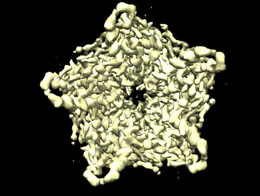
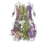
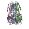
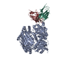
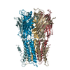
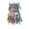
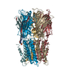
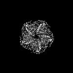
| Title | BEST1 calcium-free closed state | |||||||||
 Map data Map data | BEST1 calcium-free closed state | |||||||||
 Sample Sample |
| |||||||||
 Keywords Keywords | ion channel / calcium activated chloride channel / eukaryotic membrane protein / anion channel / transport protein / ligand gated ion channel / MEMBRANE PROTEIN | |||||||||
| Function / homology | Bestrophin-1 / Stimuli-sensing channels / Bestrophin / Bestrophin/UPF0187 / Bestrophin, RFP-TM, chloride channel / chloride channel activity / metal ion binding / membrane / Bestrophin 1 Function and homology information Function and homology information | |||||||||
| Biological species |  | |||||||||
| Method | single particle reconstruction / cryo EM / Resolution: 3.0 Å | |||||||||
 Authors Authors | Miller AN / Vaisey G | |||||||||
| Funding support |  United States, 2 items United States, 2 items
| |||||||||
 Citation Citation | Journal: Elife / Year: 2019 Title: Molecular mechanisms of gating in the calcium-activated chloride channel bestrophin. Authors: Alexandria N Miller / George Vaisey / Stephen B Long / Abstract: Bestrophin (BEST1-4) ligand-gated chloride (Cl) channels are activated by calcium (Ca). Mutation of BEST1 causes retinal disease. Partly because bestrophin channels have no sequence or structural ...Bestrophin (BEST1-4) ligand-gated chloride (Cl) channels are activated by calcium (Ca). Mutation of BEST1 causes retinal disease. Partly because bestrophin channels have no sequence or structural similarity to other ion channels, the molecular mechanisms underlying gating are unknown. Here, we present a series of cryo-electron microscopy structures of chicken BEST1, determined at 3.1 Å resolution or better, that represent the channel's principal gating states. Unlike other channels, opening of the pore is due to the repositioning of tethered pore-lining helices within a surrounding protein shell that dramatically widens a neck of the pore through a concertina of amino acid rearrangements. The neck serves as both the activation and the inactivation gate. Ca binding instigates opening of the neck through allosteric means whereas inactivation peptide binding induces closing. An aperture within the otherwise wide pore controls anion permeability. The studies define a new molecular paradigm for gating among ligand-gated ion channels. | |||||||||
| History |
|
- Structure visualization
| Movie |
Movie viewer |
|---|---|
| Structure viewer | EM map: SurfViewMolmilJmol/JSmol |
| Supplemental images |
- Downloads & links
-EMDB archive
| Map data | emd_9324.map.gz | 12 MB | EMDB map data format | |
|---|---|---|---|---|
| Header (meta data) | emd-9324-v30.xmlemd-9324.xml | 12.2 KB 12.2 KB | Display Display | EMDB header |
| Images |  emd_9324.png emd_9324.png | 187.3 KB | ||
| Filedesc metadata | emd-9324.cif.gz | 5.8 KB | ||
| Archive directory |  http://ftp.pdbj.org/pub/emdb/structures/EMD-9324ftp://ftp.pdbj.org/pub/emdb/structures/EMD-9324 http://ftp.pdbj.org/pub/emdb/structures/EMD-9324ftp://ftp.pdbj.org/pub/emdb/structures/EMD-9324 | HTTPS FTP |
-Validation report
| Summary document | emd_9324_validation.pdf.gz | 622.9 KB | Display | EMDB validaton report |
|---|---|---|---|---|
| Full document | emd_9324_full_validation.pdf.gz | 622.5 KB | Display | |
| Data in XML | emd_9324_validation.xml.gz | 5.8 KB | Display | |
| Data in CIF | emd_9324_validation.cif.gz | 6.6 KB | Display | |
| Arichive directory | https://ftp.pdbj.org/pub/emdb/validation_reports/EMD-9324ftp://ftp.pdbj.org/pub/emdb/validation_reports/EMD-9324 | HTTPS FTP |
-Related structure data
| Related structure data |  6n26MC  9321C 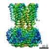 9322C 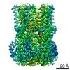 9323C 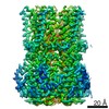 9325C 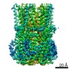 9326C 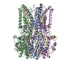 6n23C 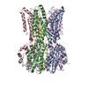 6n24C 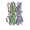 6n25C 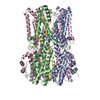 6n27C 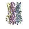 6n28C M: atomic model generated by this map C: citing same article ( |
|---|---|
| Similar structure data |

-Links
| EMDB pages | EMDB (EBI/PDBe) / EMDataResource |
|---|
-Map
| File | Download / File: emd_9324.map.gz / Format: CCP4 / Size: 12.9 MB / Type: IMAGE STORED AS FLOATING POINT NUMBER (4 BYTES) | ||||||||||||||||||||||||||||||||||||||||||||||||||||||||||||
|---|---|---|---|---|---|---|---|---|---|---|---|---|---|---|---|---|---|---|---|---|---|---|---|---|---|---|---|---|---|---|---|---|---|---|---|---|---|---|---|---|---|---|---|---|---|---|---|---|---|---|---|---|---|---|---|---|---|---|---|---|---|
| Annotation | BEST1 calcium-free closed state | ||||||||||||||||||||||||||||||||||||||||||||||||||||||||||||
| Projections & slices | Image control
Images are generated by Spider. | ||||||||||||||||||||||||||||||||||||||||||||||||||||||||||||
| Voxel size | X=Y=Z: 1.088 Å | ||||||||||||||||||||||||||||||||||||||||||||||||||||||||||||
| Density |
| ||||||||||||||||||||||||||||||||||||||||||||||||||||||||||||
| Symmetry | Space group: 1 | ||||||||||||||||||||||||||||||||||||||||||||||||||||||||||||
| Details | EMDB XML:
CCP4 map header:
| ||||||||||||||||||||||||||||||||||||||||||||||||||||||||||||
 Z (Sec.)
 Y (Row.)
 X (Col.)


-Supplemental data
- Sample components
-Entire : BEST1 calcium-free closed state
| Entire | Name: BEST1 calcium-free closed state |
|---|---|
| Components |
|
-Supramolecule #1: BEST1 calcium-free closed state
| Supramolecule | Name: BEST1 calcium-free closed state / type: complex / ID: 1 / Parent: 0 / Macromolecule list: all |
|---|---|
| Source (natural) | Organism: |
-Macromolecule #1: Bestrophin homolog
| Macromolecule | Name: Bestrophin homolog / type: protein_or_peptide / ID: 1 / Number of copies: 5 / Enantiomer: LEVO |
|---|---|
| Source (natural) | Organism: |
| Molecular weight | Theoretical: 40.761727 KDa |
| Recombinant expression | Organism:  Komagataella pastoris (fungus) Komagataella pastoris (fungus) |
| Sequence | String: TVTYTNRVAD ARLGTFSQLL LQWKGSIYKL LYSEFLIFIS LYFAISLVYR LILSESQRLM FEKLALYCNS YAELIPVSFV LGFYVSLVV SRWWAQYESI PWPDRIMNLV SCNVDGEDEY GRLLRRTLMR YSNLCSVLIL RSVSTAVYKR FPSMEHVVRA G LMTPEEHK ...String: TVTYTNRVAD ARLGTFSQLL LQWKGSIYKL LYSEFLIFIS LYFAISLVYR LILSESQRLM FEKLALYCNS YAELIPVSFV LGFYVSLVV SRWWAQYESI PWPDRIMNLV SCNVDGEDEY GRLLRRTLMR YSNLCSVLIL RSVSTAVYKR FPSMEHVVRA G LMTPEEHK KFESLNSPHN KFWIPCVWFS NLAVKARNEG RIRDSVLLQG ILNELNTLRS QCGRLYGYDW ISIPLVYTQV VT VAVYSFF LACLIGRQFL DPEKAYPGHE LDLFVPVFTF LQFFFYAGWL KVAEQLINPF GEDDDDFETN WLIDRNLQVS LMA VDEMHQ DLPILEKDLY WNEPDPQEGE EF UniProtKB: Bestrophin 1 |
-Experimental details
-Structure determination
| Method | cryo EM |
|---|---|
 Processing Processing | single particle reconstruction |
| Aggregation state | particle |
-Sample preparation
| Concentration | 5 mg/mL |
|---|---|
| Buffer | pH: 7.5 |
| Grid | Details: unspecified |
| Vitrification | Cryogen name: ETHANE / Chamber humidity: 100 % / Chamber temperature: 293 K / Instrument: FEI VITROBOT MARK IV |
- Electron microscopy
| Microscope | FEI TITAN KRIOS |
|---|---|
| Image recording | Film or detector model: GATAN K2 SUMMIT (4k x 4k) / Detector mode: SUPER-RESOLUTION / Digitization - Frames/image: 1-40 / Number grids imaged: 1 / Average exposure time: 10.0 sec. / Average electron dose: 76.0 e/Å2 |
| Electron beam | Acceleration voltage: 300 kV / Electron source:  FIELD EMISSION GUN FIELD EMISSION GUN |
| Electron optics | Illumination mode: OTHER / Imaging mode: BRIGHT FIELD |
| Sample stage | Specimen holder model: FEI TITAN KRIOS AUTOGRID HOLDER / Cooling holder cryogen: NITROGEN |
| Experimental equipment |  Model: Titan Krios / Image courtesy: FEI Company |