 Movie
Movie Controller

[English] 日本語
 Yorodumi
- PDB-8tic: Structure of human beta 1,3-N-acetylglucosaminyltransferase 2 wit... -
+ Open data

- Basic information
| Entry | Database: PDB / ID: 8tic | ||||||
|---|---|---|---|---|---|---|---|
| Title | Structure of human beta 1,3-N-acetylglucosaminyltransferase 2 with compound 1 | ||||||
 Components Components | N-acetyllactosaminide beta-1,3-N-acetylglucosaminyltransferase 2 | ||||||
 Keywords Keywords | TRANSFERASE / acetylglucosaminyltransferase / inhibitor | ||||||
| Function / homology |  Function and homology information Function and homology informationN-acetyllactosaminide beta-1,3-N-acetylglucosaminyltransferase / N-acetyllactosaminide beta-1,3-N-acetylglucosaminyltransferase activity / keratan sulfate proteoglycan biosynthetic process / poly-N-acetyllactosamine biosynthetic process / N-acetyl-beta-D-glucosaminide beta-(1,3)-galactosyltransferase activity / Keratan sulfate biosynthesis / protein O-linked glycosylation via N-acetyl-galactosamine / O-linked glycosylation of mucins / protein O-linked glycosylation / axon guidance ...N-acetyllactosaminide beta-1,3-N-acetylglucosaminyltransferase / N-acetyllactosaminide beta-1,3-N-acetylglucosaminyltransferase activity / keratan sulfate proteoglycan biosynthetic process / poly-N-acetyllactosamine biosynthetic process / N-acetyl-beta-D-glucosaminide beta-(1,3)-galactosyltransferase activity / Keratan sulfate biosynthesis / protein O-linked glycosylation via N-acetyl-galactosamine / O-linked glycosylation of mucins / protein O-linked glycosylation / axon guidance / cellular response to leukemia inhibitory factor / sensory perception of smell / Golgi membrane Similarity search - Function | ||||||
| Biological species |  Homo sapiens (human) Homo sapiens (human) | ||||||
| Method |  X-RAY DIFFRACTION / SYNCHROTRON / MOLECULAR REPLACEMENT / Resolution: 2.7 Å X-RAY DIFFRACTION / SYNCHROTRON / MOLECULAR REPLACEMENT / Resolution: 2.7 Å | ||||||
 Authors Authors | Sudom, A. / Min, X. | ||||||
| Funding support |  United States, 1items United States, 1items
| ||||||
 Citation Citation | Journal: J.Med.Chem. / Year: 2023 Title: Imidazolone as an Amide Bioisostere in the Development of beta-1,3- N -Acetylglucosaminyltransferase 2 (B3GNT2) Inhibitors. Authors: Jackson, J.J. / Siegmund, A.C. / Bai, W.J. / Reed, A.B. / Birkholz, A.B. / Campuzano, I.D.G. / Crequer-Grandhomme, A. / Hu, R. / Modak, R.V. / Sudom, A. / Javier, N. / Sanders, C. / Lo, M.C. ...Authors: Jackson, J.J. / Siegmund, A.C. / Bai, W.J. / Reed, A.B. / Birkholz, A.B. / Campuzano, I.D.G. / Crequer-Grandhomme, A. / Hu, R. / Modak, R.V. / Sudom, A. / Javier, N. / Sanders, C. / Lo, M.C. / Xie, F. / Cee, V.J. / Manzanillo, P. / Allen, J.G. | ||||||
| History |
|
- Structure visualization
| Structure viewer | Molecule: MolmilJmol/JSmol |
|---|
- Downloads & links
-Download
| PDBx/mmCIF format | 8tic.cif.gz | 286.7 KB | Display | PDBx/mmCIF format |
|---|---|---|---|---|
| PDB format | pdb8tic.ent.gz | 230.9 KB | Display | PDB format |
| PDBx/mmJSON format | 8tic.json.gz | Tree view | PDBx/mmJSON format | |
| Others |  Other downloads Other downloads |
-Validation report
| Summary document | 8tic_validation.pdf.gz | 2.7 MB | Display | wwPDB validaton report |
|---|---|---|---|---|
| Full document | 8tic_full_validation.pdf.gz | 2.7 MB | Display | |
| Data in XML | 8tic_validation.xml.gz | 50.2 KB | Display | |
| Data in CIF | 8tic_validation.cif.gz | 66.1 KB | Display | |
| Arichive directory | https://data.pdbj.org/pub/pdb/validation_reports/ti/8ticftp://data.pdbj.org/pub/pdb/validation_reports/ti/8tic | HTTPS FTP |
-Related structure data
| Related structure data |  8sz3C 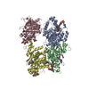 8tjcC 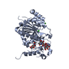 7jhkS C: citing same article ( S: Starting model for refinement |
|---|---|
| Similar structure data |
-Links
 PDBj
PDBj- Assembly
| Deposited unit | 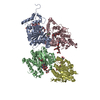
| ||||||||||
|---|---|---|---|---|---|---|---|---|---|---|---|
| 1 | 
| ||||||||||
| 2 | 
| ||||||||||
| Unit cell |
|
-Components
-Protein , 1 types, 4 molecules ABCD
| #1: Protein | Mass: 46085.898 Da / Num. of mol.: 4 Source method: isolated from a genetically manipulated source Source: (gene. exp.) Homo sapiens (human) / Gene: B3GNT2, B3GALT7, B3GNT1 / Production host:   Spodoptera frugiperda (fall armyworm) Spodoptera frugiperda (fall armyworm)References: UniProt: Q9NY97, N-acetyllactosaminide beta-1,3-N-acetylglucosaminyltransferase |
|---|
-Sugars , 2 types, 6 molecules 
| #2: Polysaccharide | alpha-D-mannopyranose-(1-3)-[alpha-D-mannopyranose-(1-6)]beta-D-mannopyranose-(1-4)-2-acetamido-2- ...alpha-D-mannopyranose-(1-3)-[alpha-D-mannopyranose-(1-6)]beta-D-mannopyranose-(1-4)-2-acetamido-2-deoxy-beta-D-glucopyranose-(1-4)-2-acetamido-2-deoxy-beta-D-glucopyranose Source method: isolated from a genetically manipulated source #3: Sugar |  Type: D-saccharide, beta linking / Mass: 221.208 Da / Num. of mol.: 2 / Source method: obtained synthetically / Formula: C8H15NO6 Type: D-saccharide, beta linking / Mass: 221.208 Da / Num. of mol.: 2 / Source method: obtained synthetically / Formula: C8H15NO6 |
|---|
-Non-polymers , 4 types, 69 molecules 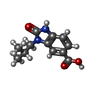


| #4: Chemical | ChemComp-FKX /  Mass: 234.251 Da / Num. of mol.: 4 / Source method: obtained synthetically / Formula: C12H14N2O3 / Feature type: SUBJECT OF INVESTIGATION Mass: 234.251 Da / Num. of mol.: 4 / Source method: obtained synthetically / Formula: C12H14N2O3 / Feature type: SUBJECT OF INVESTIGATION#5: Chemical | ChemComp-GOL /  Mass: 92.094 Da / Num. of mol.: 4 / Source method: obtained synthetically / Formula: C3H8O3 Mass: 92.094 Da / Num. of mol.: 4 / Source method: obtained synthetically / Formula: C3H8O3#6: Chemical |  Mass: 35.453 Da / Num. of mol.: 2 / Source method: obtained synthetically / Formula: Cl Mass: 35.453 Da / Num. of mol.: 2 / Source method: obtained synthetically / Formula: Cl#7: Water | ChemComp-HOH / | Mass: 18.015 Da / Num. of mol.: 59 / Source method: isolated from a natural source / Formula: H2O |
|---|
-Details
| Has ligand of interest | Y |
|---|---|
| Has protein modification | Y |
-Experimental details
-Experiment
| Experiment | Method: X-RAY DIFFRACTION / Number of used crystals: 1 |
|---|
- Sample preparation
| Crystal | Density Matthews: 2.27 Å3/Da / Density % sol: 45.87 % |
|---|---|
| Crystal grow | Temperature: 277 K / Method: vapor diffusion Details: 0.17 M sodium acetate, 0.08 M Tris pH 8.5, 25.5% PEG 4000, 15% glycerol |
-Data collection
| Diffraction | Mean temperature: 100 K / Serial crystal experiment: N |
|---|---|
| Diffraction source | Source: SYNCHROTRON / Site: ALS / Beamline: 5.0.2 / Wavelength: 1 Å |
| Detector | Type: DECTRIS PILATUS 6M / Detector: PIXEL / Date: May 11, 2018 |
| Radiation | Protocol: SINGLE WAVELENGTH / Monochromatic (M) / Laue (L): M / Scattering type: x-ray |
| Radiation wavelength | Wavelength: 1 Å / Relative weight: 1 |
| Reflection | Resolution: 2.7→29.98 Å / Num. obs: 46965 / % possible obs: 99.9 % / Redundancy: 9.6 % / Biso Wilson estimate: 44.13 Å2 / CC1/2: 0.979 / Rmerge(I) obs: 0.392 / Rpim(I) all: 0.132 / Rrim(I) all: 0.415 / Net I/σ(I): 6.2 |
| Reflection shell | Resolution: 2.7→2.79 Å / Redundancy: 10 % / Rmerge(I) obs: 2.503 / Mean I/σ(I) obs: 1.1 / Num. unique obs: 4559 / CC1/2: 0.328 / Rpim(I) all: 0.822 / Rrim(I) all: 2.639 / % possible all: 100 |
- Processing
| Software |
| |||||||||||||||||||||||||||||||||||||||||||||||||||||||||||||||||||||||||||||||||||||||||||||||||||||||||
|---|---|---|---|---|---|---|---|---|---|---|---|---|---|---|---|---|---|---|---|---|---|---|---|---|---|---|---|---|---|---|---|---|---|---|---|---|---|---|---|---|---|---|---|---|---|---|---|---|---|---|---|---|---|---|---|---|---|---|---|---|---|---|---|---|---|---|---|---|---|---|---|---|---|---|---|---|---|---|---|---|---|---|---|---|---|---|---|---|---|---|---|---|---|---|---|---|---|---|---|---|---|---|---|---|---|---|
| Refinement | Method to determine structure: MOLECULAR REPLACEMENT Starting model: 7JHK Resolution: 2.7→29.98 Å / SU ML: 0.38 / Cross valid method: FREE R-VALUE / σ(F): 1.33 / Phase error: 26.75 / Stereochemistry target values: ML
| |||||||||||||||||||||||||||||||||||||||||||||||||||||||||||||||||||||||||||||||||||||||||||||||||||||||||
| Solvent computation | Shrinkage radii: 0.9 Å / VDW probe radii: 1.1 Å / Solvent model: FLAT BULK SOLVENT MODEL | |||||||||||||||||||||||||||||||||||||||||||||||||||||||||||||||||||||||||||||||||||||||||||||||||||||||||
| Refinement step | Cycle: LAST / Resolution: 2.7→29.98 Å
| |||||||||||||||||||||||||||||||||||||||||||||||||||||||||||||||||||||||||||||||||||||||||||||||||||||||||
| Refine LS restraints |
| |||||||||||||||||||||||||||||||||||||||||||||||||||||||||||||||||||||||||||||||||||||||||||||||||||||||||
| LS refinement shell |
|
