 Movie
Movie Controller


+ Open data

- Basic information
| Entry | Database: PDB / ID: 8izb | ||||||
|---|---|---|---|---|---|---|---|
| Title | Lysophosphatidylserine receptor GPR174-Gs complex | ||||||
 Components Components |
| ||||||
 Keywords Keywords | SIGNALING PROTEIN / Complex | ||||||
| Function / homology |  Function and homology information Function and homology informationbioactive lipid receptor activity / positive regulation of Rho protein signal transduction / T cell homeostasis / PKA activation in glucagon signalling / hair follicle placode formation / centriolar satellite / mu-type opioid receptor binding / developmental growth / corticotropin-releasing hormone receptor 1 binding / intracellular transport ...bioactive lipid receptor activity / positive regulation of Rho protein signal transduction / T cell homeostasis / PKA activation in glucagon signalling / hair follicle placode formation / centriolar satellite / mu-type opioid receptor binding / developmental growth / corticotropin-releasing hormone receptor 1 binding / intracellular transport / D1 dopamine receptor binding / Hedgehog 'off' state / beta-2 adrenergic receptor binding / adenylate cyclase-activating adrenergic receptor signaling pathway / activation of adenylate cyclase activity / adenylate cyclase activator activity / trans-Golgi network membrane / G protein-coupled receptor activity / ionotropic glutamate receptor binding / insulin-like growth factor receptor binding / G-protein beta/gamma-subunit complex binding / Olfactory Signaling Pathway / Activation of the phototransduction cascade / bone development / G beta:gamma signalling through PLC beta / Presynaptic function of Kainate receptors / Thromboxane signalling through TP receptor / G-protein activation / adenylate cyclase-activating G protein-coupled receptor signaling pathway / G protein-coupled acetylcholine receptor signaling pathway / Activation of G protein gated Potassium channels / Inhibition of voltage gated Ca2+ channels via Gbeta/gamma subunits / Prostacyclin signalling through prostacyclin receptor / Glucagon signaling in metabolic regulation / G beta:gamma signalling through CDC42 / cognition / ADP signalling through P2Y purinoceptor 12 / G beta:gamma signalling through BTK / Synthesis, secretion, and inactivation of Glucagon-like Peptide-1 (GLP-1) / Sensory perception of sweet, bitter, and umami (glutamate) taste / photoreceptor disc membrane / platelet aggregation / Adrenaline,noradrenaline inhibits insulin secretion / Glucagon-type ligand receptors / Vasopressin regulates renal water homeostasis via Aquaporins / G alpha (z) signalling events / cellular response to catecholamine stimulus / Glucagon-like Peptide-1 (GLP1) regulates insulin secretion / ADORA2B mediated anti-inflammatory cytokines production / sensory perception of taste / ADP signalling through P2Y purinoceptor 1 / adenylate cyclase-activating dopamine receptor signaling pathway / G beta:gamma signalling through PI3Kgamma / cellular response to prostaglandin E stimulus / Cooperation of PDCL (PhLP1) and TRiC/CCT in G-protein beta folding / GPER1 signaling / G-protein beta-subunit binding / Inactivation, recovery and regulation of the phototransduction cascade / heterotrimeric G-protein complex / G alpha (12/13) signalling events / extracellular vesicle / sensory perception of smell / signaling receptor complex adaptor activity / Thrombin signalling through proteinase activated receptors (PARs) / GTPase binding / retina development in camera-type eye / phospholipase C-activating G protein-coupled receptor signaling pathway / Ca2+ pathway / positive regulation of cold-induced thermogenesis / G alpha (i) signalling events / fibroblast proliferation / G alpha (s) signalling events / G alpha (q) signalling events / cell population proliferation / Ras protein signal transduction / Extra-nuclear estrogen signaling / G protein-coupled receptor signaling pathway / lysosomal membrane / intracellular membrane-bounded organelle / GTPase activity / synapse / protein-containing complex binding / GTP binding / signal transduction / extracellular exosome / membrane / metal ion binding / plasma membrane / cytoplasm / cytosol Similarity search - Function | ||||||
| Biological species |  Homo sapiens (human) Homo sapiens (human)synthetic construct (others) | ||||||
| Method | ELECTRON MICROSCOPY / single particle reconstruction / cryo EM / Resolution: 3.06 Å | ||||||
 Authors Authors | Gong, W. / Liu, G. / Li, X. / Zhang, X. | ||||||
| Funding support | 1items
| ||||||
 Citation Citation | Journal: PLoS Biol / Year: 2023 Title: Structural basis for ligand recognition and signaling of the lysophosphatidylserine receptors GPR34 and GPR174. Authors: Guibing Liu / Xiu Li / Yujing Wang / Xuan Zhang / Weimin Gong /   Abstract: Lysophosphatidylserine (LysoPS) is a naturally occurring lipid mediator involved in various physiological and pathological processes especially those related to the immune system. GPR34, GPR174, and ...Lysophosphatidylserine (LysoPS) is a naturally occurring lipid mediator involved in various physiological and pathological processes especially those related to the immune system. GPR34, GPR174, and P2Y10 have been identified as the receptors for LysoPS, and its analogues have been developed as agonists or antagonists for these receptors. However, the lack of structural information hinders the drug development with novel characteristics, such as nonlipid ligands and allosteric modulators. Here, we determined the structures of human GPR34 and GPR174 in complex with LysoPS and G protein by cryo-EM. Combined with structural analysis and functional studies, we elucidated the lipid-binding modes of these receptors. By structural comparison, we identified the structural features of GPR34 and GPR174 in active state. Taken together, our findings provide insights into ligand recognition and signaling of LysoPS receptors and will facilitate the development of novel therapeutics for related inflammatory diseases and autoimmune diseases. | ||||||
| History |
|
- Structure visualization
| Structure viewer | Molecule: MolmilJmol/JSmol |
|---|
- Downloads & links
-Download
| PDBx/mmCIF format | 8izb.cif.gz | 229.7 KB | Display | PDBx/mmCIF format |
|---|---|---|---|---|
| PDB format | pdb8izb.ent.gz | 173.9 KB | Display | PDB format |
| PDBx/mmJSON format | 8izb.json.gz | Tree view | PDBx/mmJSON format | |
| Others |  Other downloads Other downloads |
-Validation report
| Summary document | 8izb_validation.pdf.gz | 1.4 MB | Display | wwPDB validaton report |
|---|---|---|---|---|
| Full document | 8izb_full_validation.pdf.gz | 1.3 MB | Display | |
| Data in XML | 8izb_validation.xml.gz | 37.8 KB | Display | |
| Data in CIF | 8izb_validation.cif.gz | 55.3 KB | Display | |
| Arichive directory | https://data.pdbj.org/pub/pdb/validation_reports/iz/8izbftp://data.pdbj.org/pub/pdb/validation_reports/iz/8izb | HTTPS FTP |
-Related structure data
| Related structure data |  35838MC  8wrbC M: map data used to model this data C: citing same article ( |
|---|---|
| Similar structure data |

-Links
 PDBj
PDBj


- Assembly
| Deposited unit | 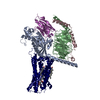
|
|---|---|
| 1 |
|
-Components
-Guanine nucleotide-binding protein ... , 3 types, 3 molecules ABC
| #1: Protein | Mass: 29048.932 Da / Num. of mol.: 1 Mutation: K25M,G49D,E50N,L63T,G226A,A249D,S252D,L272D,A366S,I372A,V375I Source method: isolated from a genetically manipulated source Source: (gene. exp.) Homo sapiens (human) / Gene: GNAS, GNAS1, GSP / Production host:   Spodoptera frugiperda (fall armyworm) / References: UniProt: P63092 Spodoptera frugiperda (fall armyworm) / References: UniProt: P63092 |
|---|---|
| #2: Protein | Mass: 40845.559 Da / Num. of mol.: 1 Source method: isolated from a genetically manipulated source Source: (gene. exp.) Homo sapiens (human) / Gene: GNB1 / Production host: Spodoptera frugiperda (fall armyworm) / References: UniProt: P62873 |
| #3: Protein | Mass: 7861.143 Da / Num. of mol.: 1 Source method: isolated from a genetically manipulated source Source: (gene. exp.) Homo sapiens (human) / Gene: GNG2 / Production host: Spodoptera frugiperda (fall armyworm) / References: UniProt: P59768 |
-Protein , 2 types, 2 molecules NR
| #4: Protein | Mass: 15140.742 Da / Num. of mol.: 1 Source method: isolated from a genetically manipulated source Source: (gene. exp.) synthetic construct (others) / Production host:  |
|---|---|
| #5: Protein | Mass: 63908.180 Da / Num. of mol.: 1 Source method: isolated from a genetically manipulated source Source: (gene. exp.) Homo sapiens (human) / Gene: GPR174, FKSG79, GPCR17 / Production host: Spodoptera frugiperda (fall armyworm) / References: UniProt: Q9BXC1 |
-Non-polymers , 3 types, 4 molecules 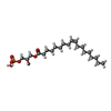


| #6: Chemical | ChemComp-UBL / [( Mass: 436.520 Da / Num. of mol.: 1 / Source method: obtained synthetically / Formula: C21H41O7P / Feature type: SUBJECT OF INVESTIGATION Mass: 436.520 Da / Num. of mol.: 1 / Source method: obtained synthetically / Formula: C21H41O7P / Feature type: SUBJECT OF INVESTIGATION |
|---|---|
| #7: Chemical | ChemComp-SER /  Type: L-peptide linking / Mass: 105.093 Da / Num. of mol.: 1 / Source method: obtained synthetically / Formula: C3H7NO3 Type: L-peptide linking / Mass: 105.093 Da / Num. of mol.: 1 / Source method: obtained synthetically / Formula: C3H7NO3 |
| #8: Chemical |  Mass: 386.654 Da / Num. of mol.: 2 / Source method: obtained synthetically / Formula: C27H46O Mass: 386.654 Da / Num. of mol.: 2 / Source method: obtained synthetically / Formula: C27H46O |
-Details
| Has ligand of interest | Y |
|---|---|
| Nonpolymer details | 1-(9Z-octadecenoyl)-sn-glycero-3-phospho-L-serine (18:1 LysoPS) is composed of UBL R 501 and SER R 502 |
-Experimental details
-Experiment
| Experiment | Method: ELECTRON MICROSCOPY |
|---|---|
| EM experiment | Aggregation state: PARTICLE / 3D reconstruction method: single particle reconstruction |
- Sample preparation
| Component | Name: LysoPS-bound GPR174 in complex with Gs and Nb35. / Type: COMPLEX / Entity ID: #1-#5 / Source: MULTIPLE SOURCES |
|---|---|
| Source (natural) | Organism: Homo sapiens (human) |
| Source (recombinant) | Organism: Spodoptera frugiperda (fall armyworm) |
| Buffer solution | pH: 7.5 |
| Specimen | Embedding applied: NO / Shadowing applied: NO / Staining applied: NO / Vitrification applied: YES |
| Vitrification | Cryogen name: ETHANE |
- Electron microscopy imaging
| Experimental equipment |  Model: Titan Krios / Image courtesy: FEI Company |
|---|---|
| Microscopy | Model: FEI TITAN KRIOS |
| Electron gun | Electron source:  FIELD EMISSION GUN / Accelerating voltage: 300 kV / Illumination mode: FLOOD BEAM FIELD EMISSION GUN / Accelerating voltage: 300 kV / Illumination mode: FLOOD BEAM |
| Electron lens | Mode: BRIGHT FIELD / Nominal defocus max: 2200 nm / Nominal defocus min: 1200 nm |
| Image recording | Electron dose: 55 e/Å2 / Film or detector model: GATAN K3 BIOQUANTUM (6k x 4k) |
- Processing
| EM software | Name: PHENIX / Version: 1.20.1_4487: / Category: model refinement | ||||||||||||||||||||||||
|---|---|---|---|---|---|---|---|---|---|---|---|---|---|---|---|---|---|---|---|---|---|---|---|---|---|
| CTF correction | Type: PHASE FLIPPING AND AMPLITUDE CORRECTION | ||||||||||||||||||||||||
| 3D reconstruction | Resolution: 3.06 Å / Resolution method: FSC 0.143 CUT-OFF / Num. of particles: 132808 / Symmetry type: POINT | ||||||||||||||||||||||||
| Refine LS restraints |
|
