 Movie
Movie Controller

[English] 日本語
 Yorodumi
- PDB-8f83: Crystal Structure of Dihydrofolate reductase (DHFR) from Mycobact... -
+ Open data

- Basic information
| Entry | Database: PDB / ID: 8f83 | |||||||||
|---|---|---|---|---|---|---|---|---|---|---|
| Title | Crystal Structure of Dihydrofolate reductase (DHFR) from Mycobacterium ulcerans Agy99 in complex with NADP and inhibitor MAM846 | |||||||||
 Components Components | Dihydrofolate reductase | |||||||||
 Keywords Keywords | OXIDOREDUCTASE/INHIBITOR / SSGCID / STRUCTURAL GENOMICS / SEATTLE STRUCTURAL GENOMICS CENTER FOR INFECTIOUS DISEASE / OXIDOREDUCTASE-OXIDOREDUCTASE INHIBITOR complex / OXIDOREDUCTASE / OXIDOREDUCTASE-INHIBITOR complex | |||||||||
| Function / homology |  Function and homology information Function and homology informationNADP+ binding / dihydrofolate metabolic process / dihydrofolate reductase / dihydrofolate reductase activity / folic acid metabolic process / tetrahydrofolate biosynthetic process / one-carbon metabolic process / cytosol Similarity search - Function | |||||||||
| Biological species |  Mycobacterium ulcerans (bacteria) Mycobacterium ulcerans (bacteria) | |||||||||
| Method |  X-RAY DIFFRACTION / SYNCHROTRON / MOLECULAR REPLACEMENT / Resolution: 1.32 Å X-RAY DIFFRACTION / SYNCHROTRON / MOLECULAR REPLACEMENT / Resolution: 1.32 Å | |||||||||
 Authors Authors | Seattle Structural Genomics Center for Infectious Disease / Seattle Structural Genomics Center for Infectious Disease (SSGCID) | |||||||||
| Funding support |  United States, 2items United States, 2items
| |||||||||
 Citation Citation | Journal: J.Med.Chem. / Year: 2024 Title: Rational Exploration of 2,4-Diaminopyrimidines as DHFR Inhibitors Active against Mycobacterium abscessus and Mycobacterium avium , Two Emerging Human Pathogens. Authors: Andrade Meirelles, M. / Almeida, V.M. / Sullivan, J.R. / de Toledo, I. / Dos Reis, C.V. / Cunha, M.R. / Zigweid, R. / Shim, A. / Sankaran, B. / Woodward, E.L. / Seibold, S. / Liu, L. / Mian, ...Authors: Andrade Meirelles, M. / Almeida, V.M. / Sullivan, J.R. / de Toledo, I. / Dos Reis, C.V. / Cunha, M.R. / Zigweid, R. / Shim, A. / Sankaran, B. / Woodward, E.L. / Seibold, S. / Liu, L. / Mian, M.R. / Battaile, K.P. / Riley, J. / Duncan, C. / Simeons, F.R.C. / Ferguson, L. / Joji, H. / Read, K.D. / Lovell, S. / Staker, B.L. / Behr, M.A. / Pilli, R.A. / Counago, R.M. | |||||||||
| History |
|
- Structure visualization
| Structure viewer | Molecule: MolmilJmol/JSmol |
|---|
- Downloads & links
-Download
| PDBx/mmCIF format | 8f83.cif.gz | 89.1 KB | Display | PDBx/mmCIF format |
|---|---|---|---|---|
| PDB format | pdb8f83.ent.gz | 63.7 KB | Display | PDB format |
| PDBx/mmJSON format | 8f83.json.gz | Tree view | PDBx/mmJSON format | |
| Others |  Other downloads Other downloads |
-Validation report
| Arichive directory | https://data.pdbj.org/pub/pdb/validation_reports/f8/8f83ftp://data.pdbj.org/pub/pdb/validation_reports/f8/8f83 | HTTPS FTP |
|---|
-Related structure data
| Related structure data | 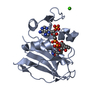 7k6cC  8f80C  8f81C  8f82C  8f84C 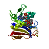 8f85C 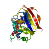 8ta0C 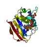 8ta1C 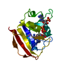 8tbrC C: citing same article ( |
|---|---|
| Similar structure data |
-Links
 PDBj
PDBj


- Assembly
| Deposited unit | 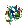
| ||||||||
|---|---|---|---|---|---|---|---|---|---|
| 1 |
| ||||||||
| Unit cell |
|
-Components
| #1: Protein | Mass: 19093.654 Da / Num. of mol.: 1 / Fragment: MyulA.01062.a.B11 / Mutation: C89S Source method: isolated from a genetically manipulated source Source: (gene. exp.) Mycobacterium ulcerans (bacteria) / Strain: Agy99 / Gene: dfrA, MUL_2179 / Plasmid: MyulA.01062.a.B11 / Production host: |
|---|---|
| #2: Chemical | ChemComp-NAP /   Mass: 743.405 Da / Num. of mol.: 1 / Source method: obtained synthetically / Formula: C21H28N7O17P3 Mass: 743.405 Da / Num. of mol.: 1 / Source method: obtained synthetically / Formula: C21H28N7O17P3 |
| #3: Chemical | ChemComp-XJZ /   Mass: 476.448 Da / Num. of mol.: 1 / Source method: obtained synthetically / Formula: C23H23F3N4O4 / Feature type: SUBJECT OF INVESTIGATION Mass: 476.448 Da / Num. of mol.: 1 / Source method: obtained synthetically / Formula: C23H23F3N4O4 / Feature type: SUBJECT OF INVESTIGATION |
| #4: Chemical | ChemComp-IMD /   Mass: 69.085 Da / Num. of mol.: 1 / Source method: obtained synthetically / Formula: C3H5N2 Mass: 69.085 Da / Num. of mol.: 1 / Source method: obtained synthetically / Formula: C3H5N2 |
| #5: Water | ChemComp-HOH /  Mass: 18.015 Da / Num. of mol.: 181 / Source method: isolated from a natural source / Formula: H2O Mass: 18.015 Da / Num. of mol.: 181 / Source method: isolated from a natural source / Formula: H2O |
| Has ligand of interest | Y |
| Has protein modification | N |
-Experimental details
-Experiment
| Experiment | Method: X-RAY DIFFRACTION / Number of used crystals: 1 |
|---|
- Sample preparation
| Crystal | Density Matthews: 1.83 Å3/Da / Density % sol: 32.62 % |
|---|---|
| Crystal grow | Temperature: 291 K / Method: vapor diffusion, sitting drop / pH: 7 Details: PACT B4: 0.1M MIB (Sodium malonate dibasic monohydrate, Imidazole, Boric acid) pH 7, MyulA.01062.a.B11.PS38558 at 10 mg/mL. Tray: Tray358B4 MAM846, Puck: PUCK HR00407 pin15, Cryo: 20% PEG 300 |
-Data collection
| Diffraction | Mean temperature: 100 K / Serial crystal experiment: N |
|---|---|
| Diffraction source | Source: SYNCHROTRON / Site: ALS / Beamline: 8.2.2 / Wavelength: 1 Å |
| Detector | Type: DECTRIS PILATUS3 S 2M / Detector: PIXEL / Date: Sep 22, 2022 |
| Radiation | Monochromator: Double Crystal Si 111 / Protocol: SINGLE WAVELENGTH / Monochromatic (M) / Laue (L): M / Scattering type: x-ray |
| Radiation wavelength | Wavelength: 1 Å / Relative weight: 1 |
| Reflection | Resolution: 1.32→35.38 Å / Num. obs: 31584 / % possible obs: 98.5 % / Redundancy: 4.3 % / CC1/2: 0.999 / Rmerge(I) obs: 0.054 / Rpim(I) all: 0.029 / Rrim(I) all: 0.062 / Χ2: 1.01 / Net I/σ(I): 13.9 / Num. measured all: 136013 |
| Reflection shell | Resolution: 1.32→1.34 Å / % possible obs: 91.7 % / Redundancy: 3.5 % / Rmerge(I) obs: 0.555 / Num. measured all: 4999 / Num. unique obs: 1442 / CC1/2: 0.734 / Rpim(I) all: 0.339 / Rrim(I) all: 0.655 / Χ2: 0.95 / Net I/σ(I) obs: 2.1 |
- Processing
| Software |
| ||||||||||||||||||||||||||||||||||||||||||||||||||||||||||||||||||||||||||||||||||||
|---|---|---|---|---|---|---|---|---|---|---|---|---|---|---|---|---|---|---|---|---|---|---|---|---|---|---|---|---|---|---|---|---|---|---|---|---|---|---|---|---|---|---|---|---|---|---|---|---|---|---|---|---|---|---|---|---|---|---|---|---|---|---|---|---|---|---|---|---|---|---|---|---|---|---|---|---|---|---|---|---|---|---|---|---|---|
| Refinement | Method to determine structure: MOLECULAR REPLACEMENT / Resolution: 1.32→35.38 Å / SU ML: 0.16 / Cross valid method: FREE R-VALUE / σ(F): 1.36 / Phase error: 21.95 / Stereochemistry target values: ML
| ||||||||||||||||||||||||||||||||||||||||||||||||||||||||||||||||||||||||||||||||||||
| Solvent computation | Shrinkage radii: 0.9 Å / VDW probe radii: 1.1 Å / Solvent model: FLAT BULK SOLVENT MODEL | ||||||||||||||||||||||||||||||||||||||||||||||||||||||||||||||||||||||||||||||||||||
| Refinement step | Cycle: LAST / Resolution: 1.32→35.38 Å
| ||||||||||||||||||||||||||||||||||||||||||||||||||||||||||||||||||||||||||||||||||||
| Refine LS restraints |
| ||||||||||||||||||||||||||||||||||||||||||||||||||||||||||||||||||||||||||||||||||||
| LS refinement shell |
|
