 Movie
Movie Controller

[English] 日本語
 Yorodumi
- PDB-8eun: MicroED structure of an Aeropyrum pernix protoglobin metallo-carb... -
+ Open data

- Basic information
| Entry | Database: PDB / ID: 8eun | |||||||||
|---|---|---|---|---|---|---|---|---|---|---|
| Title | MicroED structure of an Aeropyrum pernix protoglobin metallo-carbene complex | |||||||||
 Components Components | Protogloblin ApPgb | |||||||||
 Keywords Keywords | METAL BINDING PROTEIN / MicroED / directed evolution | |||||||||
| Function / homology | Protoglobin / Globin-sensor domain / Protoglobin / Globin/Protoglobin / Globin-like superfamily / oxygen binding / heme binding / Chem-WUF / Protogloblin ApPgb Function and homology information Function and homology information | |||||||||
| Biological species |   Aeropyrum pernix (archaea) Aeropyrum pernix (archaea) | |||||||||
| Method | ELECTRON CRYSTALLOGRAPHY / electron crystallography / cryo EM / Resolution: 2.5 Å | |||||||||
 Authors Authors | Danelius, E. / Gonen, T. / Unge, J.T. | |||||||||
| Funding support |  United States, 2items United States, 2items
| |||||||||
 Citation Citation | Journal: J Am Chem Soc / Year: 2023 Title: MicroED Structure of a Protoglobin Reactive Carbene Intermediate. Authors: Emma Danelius / Nicholas J Porter / Johan Unge / Frances H Arnold / Tamir Gonen / Abstract: Microcrystal electron diffraction (MicroED) is an emerging technique that has shown great potential for describing new chemical and biological molecular structures. Several important structures of ...Microcrystal electron diffraction (MicroED) is an emerging technique that has shown great potential for describing new chemical and biological molecular structures. Several important structures of small molecules, natural products, and peptides have been determined using methods. However, only a couple of novel protein structures have thus far been derived by MicroED. Taking advantage of recent technological advances, including higher acceleration voltage and using a low-noise detector in counting mode, we have determined the first structure of an protoglobin (Pgb) variant by MicroED using an AlphaFold2 model for phasing. The structure revealed that mutations introduced during directed evolution enhance carbene transfer activity by reorienting an α helix of Pgb into a dynamic loop, making the catalytic active site more readily accessible. After exposing the tiny crystals to the substrate, we also trapped the reactive iron-carbenoid intermediate involved in this engineered Pgb's new-to-nature activity, a challenging carbene transfer from a diazirine via a putative metallo-carbene. The bound structure discloses how an enlarged active site pocket stabilizes the carbene bound to the heme iron and, presumably, the transition state for the formation of this key intermediate. This work demonstrates that improved MicroED technology and the advancement in protein structure prediction now enable investigation of structures that was previously beyond reach. | |||||||||
| History |
|
- Structure visualization
| Structure viewer | Molecule: MolmilJmol/JSmol |
|---|
- Downloads & links
-Download
| PDBx/mmCIF format | 8eun.cif.gz | 115.1 KB | Display | PDBx/mmCIF format |
|---|---|---|---|---|
| PDB format | pdb8eun.ent.gz | 70.4 KB | Display | PDB format |
| PDBx/mmJSON format | 8eun.json.gz | Tree view | PDBx/mmJSON format | |
| Others |  Other downloads Other downloads |
-Validation report
| Arichive directory | https://data.pdbj.org/pub/pdb/validation_reports/eu/8eunftp://data.pdbj.org/pub/pdb/validation_reports/eu/8eun | HTTPS FTP |
|---|
-Related structure data
| Related structure data |  28616MC 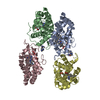 8eumC C: citing same article ( M: map data used to model this data |
|---|---|
| Similar structure data |

-Links
 PDBj
PDBj


- Assembly
| Deposited unit | 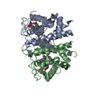
| |||||||||||||||||||||||||||||||||||||||||||||||||||||||||||
|---|---|---|---|---|---|---|---|---|---|---|---|---|---|---|---|---|---|---|---|---|---|---|---|---|---|---|---|---|---|---|---|---|---|---|---|---|---|---|---|---|---|---|---|---|---|---|---|---|---|---|---|---|---|---|---|---|---|---|---|---|
| 1 |
| |||||||||||||||||||||||||||||||||||||||||||||||||||||||||||
| Unit cell |
| |||||||||||||||||||||||||||||||||||||||||||||||||||||||||||
| Noncrystallographic symmetry (NCS) | NCS domain:
NCS domain segments: Ens-ID: ens_1
NCS oper: (Code: givenMatrix: (-0.999922402376, 0.000909120261578, 0.0124242797511), (-0.000824260945088, -0.999976311421, 0.0068335342664), (0.0124301979421, 0.0068227631518, 0.999899464987)Vector: - ...NCS oper: (Code: given Matrix: (-0.999922402376, 0.000909120261578, 0.0124242797511), Vector: |
-Components
| #1: Protein | Mass: 22718.887 Da / Num. of mol.: 2 / Mutation: C45G, W59L, Y60V, V63R, C102S, F145Q, I149L Source method: isolated from a genetically manipulated source Source: (gene. exp.) Aeropyrum pernix (archaea) / Gene: APE_0287 / Production host:  #2: Chemical | 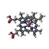  Mass: 707.618 Da / Num. of mol.: 2 / Source method: obtained synthetically / Formula: C41H39FeN4O4 / Feature type: SUBJECT OF INVESTIGATION Mass: 707.618 Da / Num. of mol.: 2 / Source method: obtained synthetically / Formula: C41H39FeN4O4 / Feature type: SUBJECT OF INVESTIGATION#3: Water | ChemComp-HOH / |  Mass: 18.015 Da / Num. of mol.: 64 / Source method: isolated from a natural source / Formula: H2O Mass: 18.015 Da / Num. of mol.: 64 / Source method: isolated from a natural source / Formula: H2OHas ligand of interest | Y | |
|---|
-Experimental details
-Experiment
| Experiment | Method: ELECTRON CRYSTALLOGRAPHY |
|---|---|
| EM experiment | Aggregation state: 3D ARRAY / 3D reconstruction method: electron crystallography |
- Sample preparation
| Component | Name: Homodimer of Aeropyrum pernix protoglobin (ApePgb) C45G, W59L, Y60V, V63R, C102S, F145Q, I149L mutant Type: COMPLEX / Entity ID: #1 / Source: RECOMBINANT |
|---|---|
| Source (natural) | Organism: Aeropyrum pernix (archaea) |
| Source (recombinant) | Organism: |
| Buffer solution | pH: 8 |
| Specimen | Conc.: 20 mg/ml / Embedding applied: NO / Shadowing applied: NO / Staining applied: NO / Vitrification applied: YES |
| Specimen support | Grid material: COPPER / Grid type: Quantifoil R2/1 |
| Vitrification | Instrument: LEICA EM GP / Cryogen name: ETHANE / Humidity: 90 % / Chamber temperature: 277 K |
-Data collection
| Experimental equipment |  Model: Titan Krios / Image courtesy: FEI Company |
|---|---|
| Microscopy | Model: FEI TITAN KRIOS |
| Electron gun | Electron source:  FIELD EMISSION GUN / Accelerating voltage: 300 kV / Illumination mode: OTHER FIELD EMISSION GUN / Accelerating voltage: 300 kV / Illumination mode: OTHER |
| Electron lens | Mode: DIFFRACTION / Nominal defocus max: 0 nm / Nominal defocus min: 0 nm / Cs: 2.7 mm / C2 aperture diameter: 50 µm / Alignment procedure: BASIC |
| Specimen holder | Cryogen: NITROGEN / Specimen holder model: FEI TITAN KRIOS AUTOGRID HOLDER / Temperature (max): 90 K / Temperature (min): 77 K |
| Image recording | Electron dose: 0.00025 e/Å2 / Detector mode: COUNTING / Film or detector model: FEI FALCON IV (4k x 4k) |
| Image scans | Sampling size: 14 µm / Width: 4096 / Height: 4096 |
| EM diffraction | Camera length: 2460 mm |
| EM diffraction shell | Resolution: 2.5→24.37 Å / Fourier space coverage: 7 % / Multiplicity: 4.3 / Num. of structure factors: 40105 / Phase residual: 29.27 ° |
| EM diffraction stats | Fourier space coverage: 70 % / High resolution: 2.5 Å / Num. of intensities measured: 40105 / Num. of structure factors: 40105 / Phase error rejection criteria: na / Rmerge: 0.32 |
| Reflection | Biso Wilson estimate: 17.58 Å2 |
- Processing
| Software |
| ||||||||||||||||||||||||||||||||||||||||||
|---|---|---|---|---|---|---|---|---|---|---|---|---|---|---|---|---|---|---|---|---|---|---|---|---|---|---|---|---|---|---|---|---|---|---|---|---|---|---|---|---|---|---|---|
| EM software | Name: PHENIX / Version: 1.20.1-4487-000 / Category: 3D reconstruction | ||||||||||||||||||||||||||||||||||||||||||
| EM 3D crystal entity | ∠α: 90 ° / ∠β: 105.42 ° / ∠γ: 90 ° / A: 58.15 Å / B: 45.89 Å / C: 71.71 Å / Space group name: P2 / Space group num: 4 | ||||||||||||||||||||||||||||||||||||||||||
| CTF correction | Type: NONE | ||||||||||||||||||||||||||||||||||||||||||
| 3D reconstruction | Resolution method: DIFFRACTION PATTERN/LAYERLINES / Symmetry type: 3D CRYSTAL | ||||||||||||||||||||||||||||||||||||||||||
| Atomic model building | B value: 20.59 / Protocol: OTHER / Space: RECIPROCAL / Target criteria: maximum likelihood | ||||||||||||||||||||||||||||||||||||||||||
| Refinement | Resolution: 2.5→24.37 Å / SU ML: 0.3157 / Cross valid method: FREE R-VALUE / σ(F): 1.34 / Phase error: 29.2747 Stereochemistry target values: GeoStd + Monomer Library + CDL v1.2
| ||||||||||||||||||||||||||||||||||||||||||
| Solvent computation | Shrinkage radii: 0.9 Å / VDW probe radii: 1.1 Å / Solvent model: FLAT BULK SOLVENT MODEL | ||||||||||||||||||||||||||||||||||||||||||
| Displacement parameters | Biso mean: 20.59 Å2 | ||||||||||||||||||||||||||||||||||||||||||
| Refinement step | Cycle: LAST / Resolution: 2.5→24.37 Å
| ||||||||||||||||||||||||||||||||||||||||||
| Refine LS restraints |
| ||||||||||||||||||||||||||||||||||||||||||
| Refine LS restraints NCS | Type: Torsion NCS / Rms dev position: 0.956942943303 Å | ||||||||||||||||||||||||||||||||||||||||||
| LS refinement shell | Refine-ID: ELECTRON CRYSTALLOGRAPHY / % reflection Rfree: 10 %
|
