 Movie
Movie Controller

[English] 日本語
 Yorodumi
- PDB-8dqp: Crystal structure of Arabidopsis thaliana COSY in complex with sc... -
+ Open data

- Basic information
| Entry | Database: PDB / ID: 8dqp | ||||||||||||
|---|---|---|---|---|---|---|---|---|---|---|---|---|---|
| Title | Crystal structure of Arabidopsis thaliana COSY in complex with scopoletin | ||||||||||||
 Components Components | Coumarin Synthase | ||||||||||||
 Keywords Keywords | PLANT PROTEIN / Coumarin Synthase / Trans/Cis Isomerase / BAHD Acyltransferase | ||||||||||||
| Function / homology |  Function and homology information Function and homology information | ||||||||||||
| Biological species |  | ||||||||||||
| Method |  X-RAY DIFFRACTION / SYNCHROTRON / MOLECULAR REPLACEMENT / Resolution: 2.48 Å X-RAY DIFFRACTION / SYNCHROTRON / MOLECULAR REPLACEMENT / Resolution: 2.48 Å | ||||||||||||
 Authors Authors | Kim, C.Y. / Mitchell, A.J. / Gutierrez, M. / Weng, J.K. | ||||||||||||
| Funding support |  United States, 3items United States, 3items
| ||||||||||||
 Citation Citation | Journal: Nat Commun / Year: 2023 Title: Emergence of a proton exchange-based isomerization and lactonization mechanism in the plant coumarin synthase COSY. Authors: Kim, C.Y. / Mitchell, A.J. / Kastner, D.W. / Albright, C.E. / Gutierrez, M.A. / Glinkerman, C.M. / Kulik, H.J. / Weng, J.K. | ||||||||||||
| History |
|
- Structure visualization
| Structure viewer | Molecule: MolmilJmol/JSmol |
|---|
- Downloads & links
-Download
| PDBx/mmCIF format | 8dqp.cif.gz | 373.3 KB | Display | PDBx/mmCIF format |
|---|---|---|---|---|
| PDB format | pdb8dqp.ent.gz | 291.7 KB | Display | PDB format |
| PDBx/mmJSON format | 8dqp.json.gz | Tree view | PDBx/mmJSON format | |
| Others |  Other downloads Other downloads |
-Validation report
| Arichive directory | https://data.pdbj.org/pub/pdb/validation_reports/dq/8dqpftp://data.pdbj.org/pub/pdb/validation_reports/dq/8dqp | HTTPS FTP |
|---|
-Related structure data
| Related structure data | 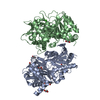 8dqoSC 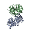 8dqqC  8dqrC S: Starting model for refinement C: citing same article ( |
|---|---|
| Similar structure data |
-Links
 PDBj
PDBj

- Assembly
| Deposited unit | 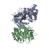
| ||||||||||||
|---|---|---|---|---|---|---|---|---|---|---|---|---|---|
| 1 |
| ||||||||||||
| Unit cell |
|
-Components
-Protein , 1 types, 2 molecules AB
| #1: Protein | Mass: 49732.254 Da / Num. of mol.: 2 Source method: isolated from a genetically manipulated source Source: (gene. exp.)  |
|---|
-Non-polymers , 6 types, 129 molecules 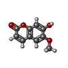


| #2: Chemical | ChemComp-T83 /  Mass: 192.168 Da / Num. of mol.: 1 / Source method: obtained synthetically / Formula: C10H8O4 / Feature type: SUBJECT OF INVESTIGATION Mass: 192.168 Da / Num. of mol.: 1 / Source method: obtained synthetically / Formula: C10H8O4 / Feature type: SUBJECT OF INVESTIGATION | ||||||||
|---|---|---|---|---|---|---|---|---|---|
| #3: Chemical |  Mass: 40.078 Da / Num. of mol.: 2 / Source method: obtained synthetically / Formula: Ca Mass: 40.078 Da / Num. of mol.: 2 / Source method: obtained synthetically / Formula: Ca#4: Chemical |  Mass: 62.068 Da / Num. of mol.: 2 / Source method: obtained synthetically / Formula: C2H6O2 Mass: 62.068 Da / Num. of mol.: 2 / Source method: obtained synthetically / Formula: C2H6O2#5: Chemical | ChemComp-PG4 / |  Mass: 194.226 Da / Num. of mol.: 1 / Source method: obtained synthetically / Formula: C8H18O5 / Comment: precipitant*YM Mass: 194.226 Da / Num. of mol.: 1 / Source method: obtained synthetically / Formula: C8H18O5 / Comment: precipitant*YM#6: Chemical | ChemComp-ACT / |  Mass: 59.044 Da / Num. of mol.: 1 / Source method: obtained synthetically / Formula: C2H3O2 Mass: 59.044 Da / Num. of mol.: 1 / Source method: obtained synthetically / Formula: C2H3O2#7: Water | ChemComp-HOH / | Mass: 18.015 Da / Num. of mol.: 122 / Source method: isolated from a natural source / Formula: H2O |
-Details
| Has ligand of interest | Y |
|---|
-Experimental details
-Experiment
| Experiment | Method: X-RAY DIFFRACTION / Number of used crystals: 1 |
|---|
- Sample preparation
| Crystal | Density Matthews: 2.34 Å3/Da / Density % sol: 47.5 % |
|---|---|
| Crystal grow | Temperature: 298.15 K / Method: vapor diffusion, hanging drop Details: 50 mM calcium acetate, 0.1M sodium cacodylate pH 6.5, 20% glycerol, 500 uM scopoletin, 5 mg/mL protein PH range: 6.5-7.5 / Temp details: Room Temperature |
-Data collection
| Diffraction | Mean temperature: 100 K / Serial crystal experiment: N |
|---|---|
| Diffraction source | Source: SYNCHROTRON / Site: APS / Beamline: 24-ID-C / Wavelength: 0.9791 Å |
| Detector | Type: DECTRIS EIGER2 X 16M / Detector: PIXEL / Date: Nov 26, 2017 |
| Radiation | Protocol: SINGLE WAVELENGTH / Monochromatic (M) / Laue (L): M / Scattering type: x-ray |
| Radiation wavelength | Wavelength: 0.9791 Å / Relative weight: 1 |
| Reflection | Resolution: 2.48→136.09 Å / Num. obs: 33466 / % possible obs: 97.46 % / Redundancy: 1.9 % / Biso Wilson estimate: 60.1 Å2 / CC1/2: 0.999 / Net I/σ(I): 10.64 |
| Reflection shell | Resolution: 2.48→2.569 Å / Redundancy: 2 % / Rmerge(I) obs: 0.7324 / Mean I/σ(I) obs: 0.89 / Num. unique obs: 3278 / CC1/2: 0.474 / CC star: 0.802 / Rpim(I) all: 0.7324 / Rrim(I) all: 1.036 / % possible all: 96.88 |
- Processing
| Software |
| ||||||||||||||||||||||||||||||||||||||||||||||||||||||||||||||||||||||||||||||||||||
|---|---|---|---|---|---|---|---|---|---|---|---|---|---|---|---|---|---|---|---|---|---|---|---|---|---|---|---|---|---|---|---|---|---|---|---|---|---|---|---|---|---|---|---|---|---|---|---|---|---|---|---|---|---|---|---|---|---|---|---|---|---|---|---|---|---|---|---|---|---|---|---|---|---|---|---|---|---|---|---|---|---|---|---|---|---|
| Refinement | Method to determine structure: MOLECULAR REPLACEMENT Starting model: 8DQO Resolution: 2.48→68.32 Å / SU ML: 0.3893 / Cross valid method: FREE R-VALUE / σ(F): 0.31 / Phase error: 36.6216 Stereochemistry target values: GeoStd + Monomer Library + CDL v1.2
| ||||||||||||||||||||||||||||||||||||||||||||||||||||||||||||||||||||||||||||||||||||
| Solvent computation | Shrinkage radii: 0.9 Å / VDW probe radii: 1.11 Å / Solvent model: FLAT BULK SOLVENT MODEL | ||||||||||||||||||||||||||||||||||||||||||||||||||||||||||||||||||||||||||||||||||||
| Refinement step | Cycle: LAST / Resolution: 2.48→68.32 Å
| ||||||||||||||||||||||||||||||||||||||||||||||||||||||||||||||||||||||||||||||||||||
| Refine LS restraints |
| ||||||||||||||||||||||||||||||||||||||||||||||||||||||||||||||||||||||||||||||||||||
| LS refinement shell |
|
