 Movie
Movie Controller

[English] 日本語
 Yorodumi
- PDB-8a3h: Cellobiose-derived imidazole complex of the endoglucanase cel5A f... -
+ Open data

- Basic information
| Entry | Database: PDB / ID: 8a3h | |||||||||
|---|---|---|---|---|---|---|---|---|---|---|
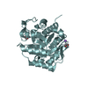
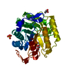
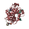
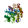
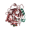
| Title | Cellobiose-derived imidazole complex of the endoglucanase cel5A from Bacillus agaradhaerens at 0.97 A resolution | |||||||||
 Components Components | PROTEIN (ENDOGLUCANASE) | |||||||||
 Keywords Keywords | HYDROLASE / CELLULOSE DEGRADATION / GLYCOSIDE HYDROLASE FAMILY 5 / ENDOGLUCANASE / TRANSITION STATE ANALOGUE / LATERAL PROTONATION | |||||||||
| Function / homology |  Function and homology information Function and homology informationcellulase / cellulase activity / cellulose catabolic process / carbohydrate binding / extracellular region Similarity search - Function | |||||||||
| Biological species |  Bacillus agaradhaerens (bacteria) Bacillus agaradhaerens (bacteria) | |||||||||
| Method |  X-RAY DIFFRACTION / SYNCHROTRON / OTHER / Resolution: 0.97 Å X-RAY DIFFRACTION / SYNCHROTRON / OTHER / Resolution: 0.97 Å | |||||||||
 Authors Authors | Varrot, A. / Schulein, M. / Pipelier, M. / Vasella, A. / Davies, G.J. | |||||||||
 Citation Citation | Journal: J.Am.Chem.Soc. / Year: 1999 Title: Lateral Protonation of a Glycosidase Inhibitor. Structure of the Bacillus agaradhaerens Cel5A in Complex with a Cellobiose-Derived Imidazole at 0.97 A Resolution Authors: Varrot, A. / Schulein, M. / Pipelier, M. / Vasella, A. / Davies, G.J. | |||||||||
| History |
|
- Structure visualization
| Structure viewer | Molecule: MolmilJmol/JSmol |
|---|
- Downloads & links
-Download
| PDBx/mmCIF format | 8a3h.cif.gz | 146.6 KB | Display | PDBx/mmCIF format |
|---|---|---|---|---|
| PDB format | pdb8a3h.ent.gz | 114.7 KB | Display | PDB format |
| PDBx/mmJSON format | 8a3h.json.gz | Tree view | PDBx/mmJSON format | |
| Others |  Other downloads Other downloads |
-Validation report
| Arichive directory | https://data.pdbj.org/pub/pdb/validation_reports/a3/8a3hftp://data.pdbj.org/pub/pdb/validation_reports/a3/8a3h | HTTPS FTP |
|---|
-Related structure data
| Similar structure data |
|---|


-Links
 PDBj
PDBj- Assembly
| Deposited unit | 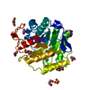
| ||||||||
|---|---|---|---|---|---|---|---|---|---|
| 1 |
| ||||||||
| Unit cell |
|
-Components
-Protein / Sugars , 2 types, 2 molecules A
| #1: Protein | Mass: 33998.023 Da / Num. of mol.: 1 / Fragment: CATALYTIC CORE DOMAIN Source method: isolated from a genetically manipulated source Source: (gene. exp.) Bacillus agaradhaerens (bacteria) / Strain: AC13 (NCIMB 40482) / Plasmid: THERRAMYL-AMYLASE PROMOTER SYSTEM / Production host: References: UniProt: P06565, UniProt: O85465*PLUS, cellulase |
|---|---|
| #4: Sugar | ChemComp-IDC / ( Type: D-saccharide / Mass: 362.332 Da / Num. of mol.: 1 / Source method: obtained synthetically / Formula: C14H22N2O9 Type: D-saccharide / Mass: 362.332 Da / Num. of mol.: 1 / Source method: obtained synthetically / Formula: C14H22N2O9 |
-Non-polymers , 4 types, 508 molecules 


| #2: Chemical | ChemComp-SO4 /  Mass: 96.063 Da / Num. of mol.: 1 / Source method: obtained synthetically / Formula: SO4 Mass: 96.063 Da / Num. of mol.: 1 / Source method: obtained synthetically / Formula: SO4 | ||
|---|---|---|---|
| #3: Chemical | ChemComp-ACT /  Mass: 59.044 Da / Num. of mol.: 1 / Source method: obtained synthetically / Formula: C2H3O2 Mass: 59.044 Da / Num. of mol.: 1 / Source method: obtained synthetically / Formula: C2H3O2 | ||
| #5: Chemical | ChemComp-GOL /  Mass: 92.094 Da / Num. of mol.: 7 / Source method: obtained synthetically / Formula: C3H8O3 Mass: 92.094 Da / Num. of mol.: 7 / Source method: obtained synthetically / Formula: C3H8O3#6: Water | ChemComp-HOH / | Mass: 18.015 Da / Num. of mol.: 499 / Source method: isolated from a natural source / Formula: H2O |
-Details
| Compound details | THE THREE FIRST RESIDUES ARE DISORDERED SO SEQUENCE STARTS AT RESIDUE SER A 4 . THIS IS THE ...THE THREE FIRST RESIDUES ARE DISORDERED |
|---|---|
| Nonpolymer details | THE IMIDAZOLE-DERIVED CELLOBIOSIDE IS BOUND IN THE -2 AND -1 SUBSITE. THE IMIDAZOLE RING SITE IN ...THE IMIDAZOLE-DERIVED CELLOBIOSI |
| Sequence details | THE FIRST 26 RESIDUES IN THE DATABASE CORRESPONDS TO THE PROSEQUENCE. OUR NUMBERING BEGINS AT THE ...THE FIRST 26 RESIDUES IN THE DATABASE CORRESPOND |
-Experimental details
-Experiment
| Experiment | Method: X-RAY DIFFRACTION / Number of used crystals: 1 |
|---|
- Sample preparation
| Crystal | Density Matthews: 2.16 Å3/Da / Density % sol: 43.14 % |
|---|---|
| Crystal grow | pH: 5.5 Details: PROTEIN CONCENTRATION 20 MG/ML, 2 M AMMONIUM SULPHATE, 100 MM SODIUM CITRATE PH 5.5, 10% GLYCEROL |
| Crystal grow | *PLUS Method: other / Details: Murshudov, G.N., (1997) Acta Cryst., D53, 240. |
-Data collection
| Diffraction | Mean temperature: 100 K |
|---|---|
| Diffraction source | Source: SYNCHROTRON / Site: EMBL/DESY, HAMBURG  / Beamline: X11 / Wavelength: 0.9076 / Beamline: X11 / Wavelength: 0.9076 |
| Detector | Date: Oct 15, 1998 |
| Radiation | Protocol: SINGLE WAVELENGTH / Monochromatic (M) / Laue (L): M / Scattering type: x-ray |
| Radiation wavelength | Wavelength: 0.9076 Å / Relative weight: 1 |
| Reflection | Resolution: 0.97→30 Å / Num. obs: 149215 / % possible obs: 93 % / Redundancy: 5.3 % / Biso Wilson estimate: 6.44 Å2 / Rmerge(I) obs: 0.032 / Rsym value: 0.032 / Net I/σ(I): 44.7 |
| Reflection shell | Resolution: 0.97→1 Å / Redundancy: 3.2 % / Rmerge(I) obs: 0.321 / Mean I/σ(I) obs: 3.2 / Rsym value: 0.321 / % possible all: 54 |
| Reflection | *PLUS % possible obs: 93 % / Redundancy: 5.4 % |
- Processing
| Software |
| ||||||||||||||||||||||||||||||||||||||||||||||||||||||||||||||||||||||||||||||||||||
|---|---|---|---|---|---|---|---|---|---|---|---|---|---|---|---|---|---|---|---|---|---|---|---|---|---|---|---|---|---|---|---|---|---|---|---|---|---|---|---|---|---|---|---|---|---|---|---|---|---|---|---|---|---|---|---|---|---|---|---|---|---|---|---|---|---|---|---|---|---|---|---|---|---|---|---|---|---|---|---|---|---|---|---|---|---|
| Refinement | Method to determine structure: OTHER / Resolution: 0.97→20 Å / Cross valid method: THROUGHOUT / σ(F): 0
| ||||||||||||||||||||||||||||||||||||||||||||||||||||||||||||||||||||||||||||||||||||
| Refinement step | Cycle: LAST / Resolution: 0.97→20 Å
| ||||||||||||||||||||||||||||||||||||||||||||||||||||||||||||||||||||||||||||||||||||
| Refine LS restraints |
| ||||||||||||||||||||||||||||||||||||||||||||||||||||||||||||||||||||||||||||||||||||
| Refinement | *PLUS σ(F): 0 / % reflection Rfree: 5 % / Rfactor obs: 0.1 / Rfactor Rfree: 0.12 / Rfactor Rwork: 0.1 | ||||||||||||||||||||||||||||||||||||||||||||||||||||||||||||||||||||||||||||||||||||
| Solvent computation | *PLUS | ||||||||||||||||||||||||||||||||||||||||||||||||||||||||||||||||||||||||||||||||||||
| Displacement parameters | *PLUS | ||||||||||||||||||||||||||||||||||||||||||||||||||||||||||||||||||||||||||||||||||||
| Refine LS restraints | *PLUS Type: p_plane_restr / Dev ideal: 0.034 |
