 Movie
Movie Controller


+ Open data

- Basic information
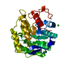
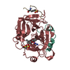
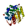
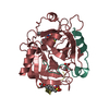
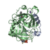
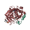
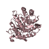
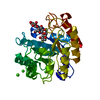
| Entry | Database: PDB / ID: 1lf1 | ||||||
|---|---|---|---|---|---|---|---|
| Title | Crystal Structure of Cel5 from Alkalophilic Bacillus sp. | ||||||
 Components Components | Cel5 | ||||||
 Keywords Keywords | HYDROLASE / Cellulose degradation | ||||||
| Function / homology |  Function and homology information Function and homology informationcellulase / cellulase activity / cellulose catabolic process / carbohydrate binding / extracellular region Similarity search - Function | ||||||
| Biological species |  | ||||||
| Method |  X-RAY DIFFRACTION / Resolution: 1.7 Å X-RAY DIFFRACTION / Resolution: 1.7 Å | ||||||
 Authors Authors | Shaw, A. / Bott, R. / Vonrhein, C. / Bricogne, G. / Power, S. / Day, A.G. | ||||||
 Citation Citation | Journal: J.Mol.Biol. / Year: 2002 Title: A novel combination of two classic catalytic schemes. Authors: Shaw, A. / Bott, R. / Vonrhein, C. / Bricogne, G. / Power, S. / Day, A.G. | ||||||
| History |
|
- Structure visualization
| Structure viewer | Molecule: MolmilJmol/JSmol |
|---|
- Downloads & links
-Download
| PDBx/mmCIF format | 1lf1.cif.gz | 70.6 KB | Display | PDBx/mmCIF format |
|---|---|---|---|---|
| PDB format | pdb1lf1.ent.gz | 52.5 KB | Display | PDB format |
| PDBx/mmJSON format | 1lf1.json.gz | Tree view | PDBx/mmJSON format | |
| Others |  Other downloads Other downloads |
-Validation report
| Arichive directory | https://data.pdbj.org/pub/pdb/validation_reports/lf/1lf1ftp://data.pdbj.org/pub/pdb/validation_reports/lf/1lf1 | HTTPS FTP |
|---|
-Related structure data
| Similar structure data |
|---|


-Links
 PDBj
PDBj- Assembly
| Deposited unit | 
| ||||||||
|---|---|---|---|---|---|---|---|---|---|
| 1 |
| ||||||||
| Unit cell |
|
-Components
| #1: Protein | Mass: 34502.590 Da / Num. of mol.: 1 Source method: isolated from a genetically manipulated source Source: (gene. exp.) Keywords: Cel5, Alkalophilic Bacillus sp. / References: UniProt: Q59232, cellulase |
|---|---|
| #2: Water | ChemComp-HOH /  Mass: 18.015 Da / Num. of mol.: 81 / Source method: isolated from a natural source / Formula: H2O Mass: 18.015 Da / Num. of mol.: 81 / Source method: isolated from a natural source / Formula: H2O |
-Experimental details
-Experiment
| Experiment | Method: X-RAY DIFFRACTION / Number of used crystals: 1 |
|---|
- Sample preparation
| Crystal | Density Matthews: 2.2 Å3/Da / Density % sol: 43.99 % |
|---|---|
| Crystal grow | Temperature: 295 K / pH: 5.5 Details: 0.5-1.0M ammonium sulfate, 200mM sodium cacodylate, pH 5.5, temperature 295K |
| Crystal grow | *PLUS Method: unknownDetails: Naki, D., (1998) Appl. Microb. Biotechnol., 49, 290. |
-Data collection
| Diffraction | Mean temperature: 295 K |
|---|---|
| Diffraction source | Source: ROTATING ANODE / Type: RIGAKU RU200 / Wavelength: 1.5418 |
| Detector | Type: RIGAKU / Detector: IMAGE PLATE |
| Radiation | Monochromator: Graphite / Protocol: SINGLE WAVELENGTH / Monochromatic (M) / Laue (L): M / Scattering type: x-ray |
| Radiation wavelength | Wavelength: 1.5418 Å / Relative weight: 1 |
| Reflection | Highest resolution: 1.7 Å / Num. obs: 32428 / % possible obs: 97.7 % / Observed criterion σ(I): 1 / Redundancy: 4.2 % / Rsym value: 0.077 / Net I/σ(I): 12.4 |
| Reflection shell | Resolution: 1.7→2 Å / Mean I/σ(I) obs: 3.61 / % possible all: 92.4 |
| Reflection | *PLUS Num. measured all: 139842 / Rmerge(I) obs: 0.077 |
- Processing
| Software | Name: SHELXL-97 / Classification: refinement | |||||||||||||||||||||||||||||||||
|---|---|---|---|---|---|---|---|---|---|---|---|---|---|---|---|---|---|---|---|---|---|---|---|---|---|---|---|---|---|---|---|---|---|---|
| Refinement | Resolution: 1.7→10 Å / Num. parameters: 9855 / Num. restraintsaints: 9682 / σ(F): 1
| |||||||||||||||||||||||||||||||||
| Refine analyze | Occupancy sum non hydrogen: 2463 | |||||||||||||||||||||||||||||||||
| Refinement step | Cycle: LAST / Resolution: 1.7→10 Å
| |||||||||||||||||||||||||||||||||
| Refine LS restraints |
| |||||||||||||||||||||||||||||||||
| Software | *PLUS Name: SHELX / Version: 97 / Classification: refinement | |||||||||||||||||||||||||||||||||
| Refinement | *PLUS Highest resolution: 1.7 Å / % reflection Rfree: 5 % / Rfactor all: 0.183 / Rfactor Rfree: 0.21 / Rfactor Rwork: 0.18 | |||||||||||||||||||||||||||||||||
| Solvent computation | *PLUS | |||||||||||||||||||||||||||||||||
| Displacement parameters | *PLUS | |||||||||||||||||||||||||||||||||
| Refine LS restraints | *PLUS
|
