+ データを開く
- 基本情報
基本情報
| 登録情報 | データベース: EMDB / ID: EMD-8852 | |||||||||
|---|---|---|---|---|---|---|---|---|---|---|
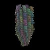
| タイトル | Cryo-EM structure of B. subtilis flagellar filaments S285P | |||||||||
 マップデータ マップデータ | Cryo-EM structure of B. subtilis flagellar filaments S285P | |||||||||
 試料 試料 |
| |||||||||
| 機能・相同性 | Flagellin, C-terminal domain / Bacterial flagellin C-terminal helical region / Flagellin / Flagellin, N-terminal domain / Bacterial flagellin N-terminal helical region / bacterial-type flagellum / structural molecule activity / extracellular region / Flagellin 機能・相同性情報 機能・相同性情報 | |||||||||
| 生物種 |  | |||||||||
| 手法 | らせん対称体再構成法 / クライオ電子顕微鏡法 / ネガティブ染色法 / 解像度: 4.5 Å | |||||||||
 データ登録者 データ登録者 | Wang F / Burrage AM / Kearns DB / Egelman EH | |||||||||
| 資金援助 |  米国, 2件 米国, 2件
| |||||||||
 引用 引用 |  ジャーナル: Nat Commun / 年: 2017 ジャーナル: Nat Commun / 年: 2017タイトル: A structural model of flagellar filament switching across multiple bacterial species. 著者: Fengbin Wang / Andrew M Burrage / Sandra Postel / Reece E Clark / Albina Orlova / Eric J Sundberg / Daniel B Kearns / Edward H Egelman /  要旨: The bacterial flagellar filament has long been studied to understand how a polymer composed of a single protein can switch between different supercoiled states with high cooperativity. Here we ...The bacterial flagellar filament has long been studied to understand how a polymer composed of a single protein can switch between different supercoiled states with high cooperativity. Here we present near-atomic resolution cryo-EM structures for flagellar filaments from both Gram-positive Bacillus subtilis and Gram-negative Pseudomonas aeruginosa. Seven mutant flagellar filaments in B. subtilis and two in P. aeruginosa capture two different states of the filament. These reliable atomic models of both states reveal conserved molecular interactions in the interior of the filament among B. subtilis, P. aeruginosa and Salmonella enterica. Using the detailed information about the molecular interactions in two filament states, we successfully predict point mutations that shift the equilibrium between those two states. Further, we observe the dimerization of P. aeruginosa outer domains without any perturbation of the conserved interior of the filament. Our results give new insights into how the flagellin sequence has been "tuned" over evolution.Bacterial flagellar filaments are composed almost entirely of a single protein-flagellin-which can switch between different supercoiled states in a highly cooperative manner. Here the authors present near-atomic resolution cryo-EM structures of nine flagellar filaments, and begin to shed light on the molecular basis of filament switching. | |||||||||
| 履歴 |
|
- 構造の表示
構造の表示
| ムービー |
 ムービービューア ムービービューア |
|---|---|
| 構造ビューア | EMマップ:  SurfView SurfView Molmil Molmil Jmol/JSmol Jmol/JSmol |
| 添付画像 |
- ダウンロードとリンク
-EMDBアーカイブ
| マップデータ |  emd_8852.map.gz emd_8852.map.gz | 129.9 MB |  EMDBマップデータ形式 EMDBマップデータ形式 | |
|---|---|---|---|---|
| ヘッダ (付随情報) |  emd-8852-v30.xml emd-8852-v30.xml emd-8852.xml emd-8852.xml | 13.6 KB 13.6 KB | 表示 表示 |  EMDBヘッダ EMDBヘッダ |
| 画像 |  emd_8852.png emd_8852.png | 108.3 KB | ||
| アーカイブディレクトリ |  http://ftp.pdbj.org/pub/emdb/structures/EMD-8852 http://ftp.pdbj.org/pub/emdb/structures/EMD-8852 ftp://ftp.pdbj.org/pub/emdb/structures/EMD-8852 ftp://ftp.pdbj.org/pub/emdb/structures/EMD-8852 | HTTPS FTP |
-検証レポート
| 文書・要旨 |  emd_8852_validation.pdf.gz emd_8852_validation.pdf.gz | 396.9 KB | 表示 |  EMDB検証レポート EMDB検証レポート |
|---|---|---|---|---|
| 文書・詳細版 |  emd_8852_full_validation.pdf.gz emd_8852_full_validation.pdf.gz | 396.4 KB | 表示 | |
| XML形式データ |  emd_8852_validation.xml.gz emd_8852_validation.xml.gz | 4.6 KB | 表示 | |
| CIF形式データ |  emd_8852_validation.cif.gz emd_8852_validation.cif.gz | 5.1 KB | 表示 | |
| アーカイブディレクトリ |  https://ftp.pdbj.org/pub/emdb/validation_reports/EMD-8852 https://ftp.pdbj.org/pub/emdb/validation_reports/EMD-8852 ftp://ftp.pdbj.org/pub/emdb/validation_reports/EMD-8852 ftp://ftp.pdbj.org/pub/emdb/validation_reports/EMD-8852 | HTTPS FTP |
-関連構造データ
| 関連構造データ |  5wjyMC  8847C  8848C  8849C  8850C  8851C  8853C  8855C  8856C  5wjtC  5wjuC  5wjvC  5wjwC  5wjxC  5wjzC  5wk5C  5wk6C C: 同じ文献を引用 ( M: このマップから作成された原子モデル |
|---|---|
| 類似構造データ |
- リンク
リンク
| EMDBのページ |  EMDB (EBI/PDBe) / EMDB (EBI/PDBe) /  EMDataResource EMDataResource |
|---|
- マップ
マップ
| ファイル |  ダウンロード / ファイル: emd_8852.map.gz / 形式: CCP4 / 大きさ: 732.4 MB / タイプ: IMAGE STORED AS FLOATING POINT NUMBER (4 BYTES) ダウンロード / ファイル: emd_8852.map.gz / 形式: CCP4 / 大きさ: 732.4 MB / タイプ: IMAGE STORED AS FLOATING POINT NUMBER (4 BYTES) | ||||||||||||||||||||||||||||||||||||||||||||||||||||||||||||
|---|---|---|---|---|---|---|---|---|---|---|---|---|---|---|---|---|---|---|---|---|---|---|---|---|---|---|---|---|---|---|---|---|---|---|---|---|---|---|---|---|---|---|---|---|---|---|---|---|---|---|---|---|---|---|---|---|---|---|---|---|---|
| 注釈 | Cryo-EM structure of B. subtilis flagellar filaments S285P | ||||||||||||||||||||||||||||||||||||||||||||||||||||||||||||
| 投影像・断面図 | 画像のコントロール
画像は Spider により作成 これらの図は立方格子座標系で作成されたものです | ||||||||||||||||||||||||||||||||||||||||||||||||||||||||||||
| ボクセルのサイズ | X=Y=Z: 0.525 Å | ||||||||||||||||||||||||||||||||||||||||||||||||||||||||||||
| 密度 |
| ||||||||||||||||||||||||||||||||||||||||||||||||||||||||||||
| 対称性 | 空間群: 1 | ||||||||||||||||||||||||||||||||||||||||||||||||||||||||||||
| 詳細 | EMDB XML:
CCP4マップ ヘッダ情報:
| ||||||||||||||||||||||||||||||||||||||||||||||||||||||||||||
-添付データ
- 試料の構成要素
-全体 : Bacillus subtilis flagella filament
| 全体 | 名称: Bacillus subtilis flagella filament |
|---|---|
| 要素 |
|
-超分子 #1: Bacillus subtilis flagella filament
| 超分子 | 名称: Bacillus subtilis flagella filament / タイプ: complex / ID: 1 / 親要素: 0 / 含まれる分子: all |
|---|---|
| 由来(天然) | 生物種:  |
| 組換発現 | 生物種:  |
-分子 #1: Flagellin
| 分子 | 名称: Flagellin / タイプ: protein_or_peptide / ID: 1 / コピー数: 41 / 光学異性体: LEVO |
|---|---|
| 由来(天然) | 生物種:  |
| 分子量 | 理論値: 32.67132 KDa |
| 組換発現 | 生物種:  |
| 配列 | 文字列: MRINHNIAAL NTLNRLSSNN SASQKNMEKL SSGLRINRAG DDAAGLAISE KMRGQIRGLE MASKNSQDGI SLIQTAEGAL TETHAILQR VRELVVQAGN TGTQDKATDL QSIQDEISAL TDEIDGISNR TEFNGKKLLD GTYKVDTATP ANQKNLVFQI G ANATQQIS ...文字列: MRINHNIAAL NTLNRLSSNN SASQKNMEKL SSGLRINRAG DDAAGLAISE KMRGQIRGLE MASKNSQDGI SLIQTAEGAL TETHAILQR VRELVVQAGN TGTQDKATDL QSIQDEISAL TDEIDGISNR TEFNGKKLLD GTYKVDTATP ANQKNLVFQI G ANATQQIS VNIEDMGADA LGIKEADGSI AALHSVNDLD VTKFADNAAD CADIGFDAQL KVVDEAINQV SSQRAKLGAV QN RLEHTIN NLSASGENLT AAESRIRDVD MAKEMSEFTK NNILSQAPQA MLAQANQQPQ NVLQLLR |
-実験情報
-構造解析
| 手法 | ネガティブ染色法, クライオ電子顕微鏡法 |
|---|---|
 解析 解析 | らせん対称体再構成法 |
| 試料の集合状態 | filament |
- 試料調製
試料調製
| 濃度 | 0.1 mg/mL |
|---|---|
| 緩衝液 | pH: 6.8 / 詳細: Imidazole buffer |
| 染色 | タイプ: NEGATIVE / 材質: negative stain |
| 凍結 | 凍結剤: ETHANE / チャンバー内湿度: 90 % / 装置: FEI VITROBOT MARK IV |
- 電子顕微鏡法
| 顕微鏡 | FEI TITAN KRIOS |
|---|---|
| 撮影 | フィルム・検出器のモデル: FEI FALCON II (4k x 4k) 検出モード: INTEGRATING / 平均露光時間: 2.0 sec. / 平均電子線量: 20.0 e/Å2 詳細: Images were stored containing seven parts, where each part represented a set of frames corresponding to a dose of ~20 electrons per Angstrom^2. The full dose image stack was used for the ...詳細: Images were stored containing seven parts, where each part represented a set of frames corresponding to a dose of ~20 electrons per Angstrom^2. The full dose image stack was used for the estimation of the CTF as well as for boxing filaments. Only the first two parts were used for the reconstruction (~5 electrons per Angstrom^2). |
| 電子線 | 加速電圧: 300 kV / 電子線源:  FIELD EMISSION GUN FIELD EMISSION GUN |
| 電子光学系 | 照射モード: FLOOD BEAM / 撮影モード: BRIGHT FIELD |
| 実験機器 |  モデル: Titan Krios / 画像提供: FEI Company |
- 画像解析
画像解析
| 最終 再構成 | 想定した対称性 - らせんパラメータ - Δz: 4.72 Å 想定した対称性 - らせんパラメータ - ΔΦ: 65.3 ° 想定した対称性 - らせんパラメータ - 軸対称性: C1 (非対称) アルゴリズム: BACK PROJECTION / 解像度のタイプ: BY AUTHOR / 解像度: 4.5 Å / 解像度の算出法: OTHER / ソフトウェア - 名称: SPIDER / 詳細: model-map FSC 0.38 cut-off / 使用した粒子像数: 55403 |
|---|---|
| CTF補正 | ソフトウェア - 名称: CTFFIND3 |
| 初期モデル | モデルのタイプ: OTHER / 詳細: featureless cylinder |
| 最終 角度割当 | タイプ: NOT APPLICABLE / ソフトウェア - 名称: SPIDER |
-原子モデル構築 1
| 精密化 | 空間: REAL |
|---|---|
| 得られたモデル |  PDB-5wjy: |
 ムービー
ムービー コントローラー


 Z (Sec.)
 Y (Row.)
 X (Col.)


