 Movie
Movie Controller

[English] 日本語
 Yorodumi
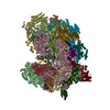
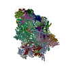
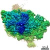
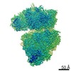
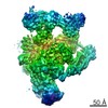
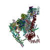
- EMDB-8190: m48S late-stage initiation complex, purified from rabbit reticulo... -
+ Open data

- Basic information
| Entry | Database: EMDB / ID: EMD-8190 | |||||||||
|---|---|---|---|---|---|---|---|---|---|---|
| Title | m48S late-stage initiation complex, purified from rabbit reticulocytes lysates, displaying eIF2 ternary complex and eIF3 i and g subunits relocated to the intersubunit face | |||||||||
 Map data Map data | None | |||||||||
 Sample Sample |
| |||||||||
 Keywords Keywords | eukaryotic translation initiation / ribosome / eIF3 peripheral subunits / cryo-EM / translation | |||||||||
| Function / homology |  Function and homology information Function and homology informationviral translational termination-reinitiation / eukaryotic translation initiation factor 3 complex, eIF3m / eukaryotic translation initiation factor 2 complex / formation of cytoplasmic translation initiation complex / eukaryotic translation initiation factor 3 complex / eukaryotic 43S preinitiation complex / formation of translation preinitiation complex / eukaryotic 48S preinitiation complex / cellular response to chemical stress / protein-synthesizing GTPase ...viral translational termination-reinitiation / eukaryotic translation initiation factor 3 complex, eIF3m / eukaryotic translation initiation factor 2 complex / formation of cytoplasmic translation initiation complex / eukaryotic translation initiation factor 3 complex / eukaryotic 43S preinitiation complex / formation of translation preinitiation complex / eukaryotic 48S preinitiation complex / cellular response to chemical stress / protein-synthesizing GTPase / Formation of the ternary complex, and subsequently, the 43S complex / laminin receptor activity / Ribosomal scanning and start codon recognition / Translation initiation complex formation / Formation of a pool of free 40S subunits / ubiquitin ligase inhibitor activity / 90S preribosome / positive regulation of signal transduction by p53 class mediator / GTP hydrolysis and joining of the 60S ribosomal subunit / L13a-mediated translational silencing of Ceruloplasmin expression / regulation of translational fidelity / phagocytic cup / translation regulator activity / gastrulation / rough endoplasmic reticulum / ribosomal small subunit export from nucleus / laminin binding / translation initiation factor activity / MDM2/MDM4 family protein binding / class I DNA-(apurinic or apyrimidinic site) endonuclease activity / response to endoplasmic reticulum stress / positive regulation of apoptotic signaling pathway / DNA-(apurinic or apyrimidinic site) lyase / cytosolic ribosome / maturation of LSU-rRNA from tricistronic rRNA transcript (SSU-rRNA, 5.8S rRNA, LSU-rRNA) / maturation of SSU-rRNA from tricistronic rRNA transcript (SSU-rRNA, 5.8S rRNA, LSU-rRNA) / maturation of SSU-rRNA / translational initiation / small-subunit processome / spindle / cytoplasmic stress granule / rRNA processing / rhythmic process / regulation of translation / ribosomal small subunit assembly / virus receptor activity / ribosome binding / ribosomal small subunit biogenesis / small ribosomal subunit / small ribosomal subunit rRNA binding / cytosolic small ribosomal subunit / cytosolic large ribosomal subunit / perikaryon / cytoplasmic translation / cell differentiation / tRNA binding / mitochondrial inner membrane / postsynaptic density / rRNA binding / structural constituent of ribosome / translation / ribonucleoprotein complex / cell division / DNA repair / mRNA binding / GTPase activity / apoptotic process / centrosome / dendrite / synapse / nucleolus / GTP binding / perinuclear region of cytoplasm / endoplasmic reticulum / DNA-templated transcription / DNA binding / RNA binding / zinc ion binding / membrane / metal ion binding / nucleus / plasma membrane / cytosol / cytoplasm Similarity search - Function | |||||||||
| Biological species |   Homo sapiens (human) Homo sapiens (human) | |||||||||
| Method | single particle reconstruction / cryo EM / Resolution: 5.8 Å | |||||||||
 Authors Authors | Simonetti A / Brito Querido J / Myasnikov AG / Mancera-Martinez E / Renaud A / Kuhn L / Hashem Y | |||||||||
| Funding support |  France, 2 items France, 2 items
| |||||||||
 Citation Citation | Journal: Mol Cell / Year: 2016 Title: eIF3 Peripheral Subunits Rearrangement after mRNA Binding and Start-Codon Recognition. Authors: Angelita Simonetti / Jailson Brito Querido / Alexander G Myasnikov / Eder Mancera-Martinez / Adeline Renaud / Lauriane Kuhn / Yaser Hashem / Abstract: mRNA translation initiation in eukaryotes requires the cooperation of a dozen eukaryotic initiation factors (eIFs) forming several complexes, which leads to mRNA attachment to the small ribosomal ...mRNA translation initiation in eukaryotes requires the cooperation of a dozen eukaryotic initiation factors (eIFs) forming several complexes, which leads to mRNA attachment to the small ribosomal 40S subunit, mRNA scanning for start codon, and accommodation of initiator tRNA at the 40S P site. eIF3, composed of 13 subunits, 8 core (a, c, e, f, h, l, k, and m) and 5 peripheral (b, d, g, i, and j), plays a central role during this process. Here we report a cryo-electron microscopy structure of a mammalian 48S initiation complex at 5.8 Å resolution. It shows the relocation of subunits eIF3i and eIF3g to the 40S intersubunit face on the GTPase binding site, at a late stage in initiation. On the basis of a previous study, we demonstrate the relocation of eIF3b to the 40S intersubunit face, binding below the eIF2-Met-tRNAi(Met) ternary complex upon mRNA attachment. Our analysis reveals the deep rearrangement of eIF3 and unravels the molecular mechanism underlying eIF3 function in mRNA scanning and timing of ribosomal subunit joining. | |||||||||
| History |
|
- Structure visualization
| Movie |
Movie viewer |
|---|---|
| Structure viewer | EM map: SurfViewMolmilJmol/JSmol |
| Supplemental images |

- Downloads & links
-EMDB archive
| Map data | emd_8190.map.gz | 28.6 MB | EMDB map data format | |
|---|---|---|---|---|
| Header (meta data) | emd-8190-v30.xmlemd-8190.xml | 66.9 KB 66.9 KB | Display Display | EMDB header |
| Images | 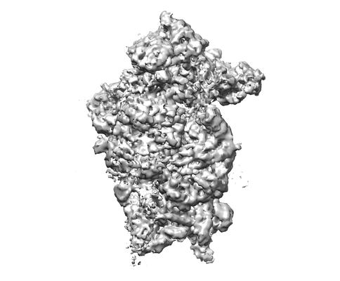 emd_8190.png emd_8190.png | 54.3 KB | ||
| Filedesc metadata | emd-8190.cif.gz | 13.3 KB | ||
| Archive directory |  http://ftp.pdbj.org/pub/emdb/structures/EMD-8190ftp://ftp.pdbj.org/pub/emdb/structures/EMD-8190 http://ftp.pdbj.org/pub/emdb/structures/EMD-8190ftp://ftp.pdbj.org/pub/emdb/structures/EMD-8190 | HTTPS FTP |
-Related structure data
| Related structure data | 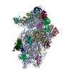 5k0yMC 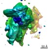 8195C  5k1hC M: atomic model generated by this map C: citing same article ( |
|---|---|
| Similar structure data |
-Links
| EMDB pages | EMDB (EBI/PDBe) / EMDataResource |
|---|---|
| Related items in Molecule of the Month |


-Map
| File | Download / File: emd_8190.map.gz / Format: CCP4 / Size: 30.5 MB / Type: IMAGE STORED AS FLOATING POINT NUMBER (4 BYTES) | ||||||||||||||||||||||||||||||||||||||||||||||||||||||||||||||||||||
|---|---|---|---|---|---|---|---|---|---|---|---|---|---|---|---|---|---|---|---|---|---|---|---|---|---|---|---|---|---|---|---|---|---|---|---|---|---|---|---|---|---|---|---|---|---|---|---|---|---|---|---|---|---|---|---|---|---|---|---|---|---|---|---|---|---|---|---|---|---|
| Annotation | None | ||||||||||||||||||||||||||||||||||||||||||||||||||||||||||||||||||||
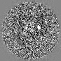
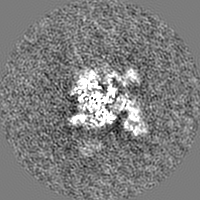
| Projections & slices | Image control
Images are generated by Spider. | ||||||||||||||||||||||||||||||||||||||||||||||||||||||||||||||||||||
| Voxel size | X=Y=Z: 2.2 Å | ||||||||||||||||||||||||||||||||||||||||||||||||||||||||||||||||||||
| Density |
| ||||||||||||||||||||||||||||||||||||||||||||||||||||||||||||||||||||
| Symmetry | Space group: 1 | ||||||||||||||||||||||||||||||||||||||||||||||||||||||||||||||||||||
| Details | EMDB XML:
CCP4 map header:
| ||||||||||||||||||||||||||||||||||||||||||||||||||||||||||||||||||||
 Z (Sec.)
 Y (Row.)
 X (Col.)


-Supplemental data
- Sample components
+Entire : m48S late-stage initiation complex, purified from rabbit reticulo...
+Supramolecule #1: m48S late-stage initiation complex, purified from rabbit reticulo...
+Macromolecule #1: tRNA
+Macromolecule #2: 18S ribosomal RNA
+Macromolecule #3: mRNA
+Macromolecule #4: Eukaryotic translation initiation factor 2 subunit 1
+Macromolecule #5: ribosomal protein uS17
+Macromolecule #6: ribosomal protein uS9
+Macromolecule #7: 40S ribosomal protein S4
+Macromolecule #8: ribosomal protein uS14
+Macromolecule #9: Ribosomal protein S9 (Predicted)
+Macromolecule #10: ribosomal protein uS13
+Macromolecule #11: Eukaryotic translation initiation factor 3 subunit G
+Macromolecule #12: Eukaryotic translation initiation factor 3 subunit G
+Macromolecule #13: ribosomal protein uS12
+Macromolecule #14: ribosomal protein eS19
+Macromolecule #15: eukaryotic initiation factor 2 Gamma subunit (eIF2-Gamma)
+Macromolecule #16: Eukaryotic translation initiation factor 3 subunit I
+Macromolecule #17: ribosomal protein uS7
+Macromolecule #18: ribosomal protein eS30
+Macromolecule #19: ribosomal protein eS25
+Macromolecule #20: ribosomal protein eS7
+Macromolecule #21: 40S ribosomal protein S27
+Macromolecule #22: ribosomal protein uS15
+Macromolecule #23: ribosomal protein uS8
+Macromolecule #24: 40S ribosomal protein S21
+Macromolecule #25: ribosomal protein uS5
+Macromolecule #26: eukaryotic initiation factor 2 subunit Beta (eIF2-Beta)
+Macromolecule #27: ribosomal protein eS17
+Macromolecule #28: ribosomal protein uS2
+Macromolecule #29: ribosomal protein uS3
+Macromolecule #30: ribosomal protein uS10
+Macromolecule #31: ribosomal protein eS1
+Macromolecule #32: ribosomal protein uS11
+Macromolecule #33: ribosomal protein eS26
+Macromolecule #34: ribosomal protein eS28
+Macromolecule #35: ribosomal protein RACK1
+Macromolecule #36: ribosomal protein uS19
+Macromolecule #37: 40S ribosomal protein S8
+Macromolecule #38: ribosomal protein eS31
+Macromolecule #39: 40S ribosomal protein S6
+Macromolecule #40: 40S ribosomal protein S12
+Macromolecule #41: 40S ribosomal protein S24
+Macromolecule #42: ribosomal protein eS10
-Experimental details
-Structure determination
| Method | cryo EM |
|---|---|
 Processing Processing | single particle reconstruction |
| Aggregation state | particle |
-Sample preparation
| Concentration | 0.5 mg/mL |
|---|---|
| Buffer | pH: 7.6 |
| Grid | Model: Quantifoil / Material: COPPER / Mesh: 400 / Support film - Material: CARBON / Support film - topology: HOLEY / Pretreatment - Type: GLOW DISCHARGE / Pretreatment - Time: 25 sec. / Pretreatment - Atmosphere: AIR |
| Vitrification | Cryogen name: ETHANE / Chamber humidity: 100 % / Chamber temperature: 277 K / Instrument: FEI VITROBOT MARK IV |
- Electron microscopy
| Microscope | FEI TITAN KRIOS |
|---|---|
| Image recording | Film or detector model: FEI FALCON II (4k x 4k) / Detector mode: INTEGRATING / Digitization - Frames/image: 2-8 / Number grids imaged: 1 / Number real images: 5700 / Average exposure time: 1.5 sec. / Average electron dose: 24.0 e/Å2 |
| Electron beam | Acceleration voltage: 300 kV / Electron source:  FIELD EMISSION GUN FIELD EMISSION GUN |
| Electron optics | C2 aperture diameter: 100.0 µm / Illumination mode: FLOOD BEAM / Imaging mode: BRIGHT FIELD / Cs: 0.01 mm / Nominal defocus max: 4.5 µm / Nominal defocus min: 0.8 µm / Nominal magnification: 59000 |
| Sample stage | Specimen holder model: FEI TITAN KRIOS AUTOGRID HOLDER / Cooling holder cryogen: NITROGEN |
| Experimental equipment |  Model: Titan Krios / Image courtesy: FEI Company |
+Image processing

-Atomic model buiding 1
| Initial model | PDB ID: Chain - Source name: PDB / Chain - Initial model type: experimental model |
|---|---|
| Refinement | Protocol: FLEXIBLE FIT |
| Output model | PDB-5k0y: |
