 Movie
Movie Controller

[English] 日本語
 Yorodumi
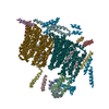
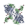
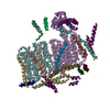
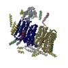
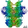
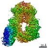
- PDB-7txz: Nipah Virus attachment (G) glycoprotein ectodomain in complex wit... -
+ Open data

- Basic information
| Entry | Database: PDB / ID: 7txz | ||||||
|---|---|---|---|---|---|---|---|
| Title | Nipah Virus attachment (G) glycoprotein ectodomain in complex with nAH1.3 neutralizing antibody Fab fragment (local refinement of the distal region) | ||||||
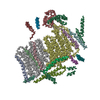
 Components Components |
| ||||||
 Keywords Keywords | VIRAL PROTEIN / NiV / NiVG / attachment protein / neutralizing antibodies / Structural Genomics / Seattle Structural Genomics Center for Infectious Disease / SSGCID | ||||||
| Function / homology |  Function and homology information Function and homology informationmembrane fusion involved in viral entry into host cell / exo-alpha-sialidase activity / clathrin-dependent endocytosis of virus by host cell / host cell surface receptor binding / viral envelope / virion attachment to host cell / host cell plasma membrane / virion membrane / identical protein binding / membrane Similarity search - Function | ||||||
| Biological species |  Nipah henipavirus Nipah henipavirus | ||||||
| Method | ELECTRON MICROSCOPY / single particle reconstruction / cryo EM / Resolution: 3.2 Å | ||||||
 Authors Authors | Wang, Z.Q. / Seattle Structural Genomics Center for Infectious Disease (SSGCID) / Veesler, D. | ||||||
| Funding support |  United States, 1items United States, 1items
| ||||||
 Citation Citation | Journal: Science / Year: 2022 Title: Architecture and antigenicity of the Nipah virus attachment glycoprotein. Authors: Zhaoqian Wang / Moushimi Amaya / Amin Addetia / Ha V Dang / Gabriella Reggiano / Lianying Yan / Andrew C Hickey / Frank DiMaio / Christopher C Broder / David Veesler / Abstract: Nipah virus (NiV) and Hendra virus (HeV) are zoonotic henipaviruses (HNVs) responsible for outbreaks of encephalitis and respiratory illness. The entry of HNVs into host cells requires the attachment ...Nipah virus (NiV) and Hendra virus (HeV) are zoonotic henipaviruses (HNVs) responsible for outbreaks of encephalitis and respiratory illness. The entry of HNVs into host cells requires the attachment (G) and fusion (F) glycoproteins, which are the main targets of antibody responses. To understand viral infection and host immunity, we determined a cryo-electron microscopy structure of the NiV G homotetrameric ectodomain in complex with the nAH1.3 broadly neutralizing antibody Fab fragment. We show that a cocktail of two nonoverlapping G-specific antibodies neutralizes NiV and HeV synergistically and limits the emergence of escape mutants. Analysis of polyclonal serum antibody responses elicited by vaccination of macaques with NiV G indicates that the receptor binding head domain is immunodominant. These results pave the way for implementing multipronged therapeutic strategies against these deadly pathogens. | ||||||
| History |
|
- Structure visualization
| Movie |
Movie viewer |
|---|---|
| Structure viewer | Molecule: MolmilJmol/JSmol |
- Downloads & links
-Download
| PDBx/mmCIF format | 7txz.cif.gz | 321.4 KB | Display | PDBx/mmCIF format |
|---|---|---|---|---|
| PDB format | pdb7txz.ent.gz | 238.2 KB | Display | PDB format |
| PDBx/mmJSON format | 7txz.json.gz | Tree view | PDBx/mmJSON format | |
| Others |  Other downloads Other downloads |
-Validation report
| Summary document | 7txz_validation.pdf.gz | 2.3 MB | Display | wwPDB validaton report |
|---|---|---|---|---|
| Full document | 7txz_full_validation.pdf.gz | 2.3 MB | Display | |
| Data in XML | 7txz_validation.xml.gz | 53.3 KB | Display | |
| Data in CIF | 7txz_validation.cif.gz | 79.7 KB | Display | |
| Arichive directory | https://data.pdbj.org/pub/pdb/validation_reports/tx/7txzftp://data.pdbj.org/pub/pdb/validation_reports/tx/7txz | HTTPS FTP |
-Related structure data
| Related structure data |  26162MC  7ty0C M: map data used to model this data C: citing same article ( |
|---|---|
| Similar structure data |


-Links
 PDBj
PDBj


- Assembly
| Deposited unit | 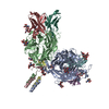
|
|---|---|
| 1 |
|
-Components
-Protein , 1 types, 4 molecules ABDC
| #1: Protein | Mass: 60358.535 Da / Num. of mol.: 4 / Fragment: Ectodomain Source method: isolated from a genetically manipulated source Source: (gene. exp.) Nipah henipavirus / Production host:  Homo sapiens (human) / References: UniProt: Q9IH62 Homo sapiens (human) / References: UniProt: Q9IH62 |
|---|
-Antibody , 2 types, 4 molecules EHFL
| #2: Antibody | Mass: 50643.941 Da / Num. of mol.: 2 Source method: isolated from a genetically manipulated source Source: (gene. exp.) Homo sapiens (human)#3: Antibody | Mass: 23947.473 Da / Num. of mol.: 2 Source method: isolated from a genetically manipulated source Source: (gene. exp.) Homo sapiens (human) |
|---|
-Sugars , 3 types, 14 molecules 
| #4: Polysaccharide | 2-acetamido-2-deoxy-beta-D-glucopyranose-(1-4)-2-acetamido-2-deoxy-beta-D-glucopyranose Source method: isolated from a genetically manipulated source #5: Polysaccharide | Source method: isolated from a genetically manipulated source #6: Sugar | ChemComp-NAG / |  Type: D-saccharide, beta linking / Mass: 221.208 Da / Num. of mol.: 1 / Source method: obtained synthetically / Formula: C8H15NO6 Type: D-saccharide, beta linking / Mass: 221.208 Da / Num. of mol.: 1 / Source method: obtained synthetically / Formula: C8H15NO6 |
|---|
-Details
| Has ligand of interest | N |
|---|---|
| Has protein modification | Y |
-Experimental details
-Experiment
| Experiment | Method: ELECTRON MICROSCOPY |
|---|---|
| EM experiment | Aggregation state: PARTICLE / 3D reconstruction method: single particle reconstruction |
- Sample preparation
| Component | Name: Nipah Virus attachment protein ectodomain in complex with nAH1.3 neutralizing antibody Fab fragment Type: COMPLEX / Entity ID: #1-#3 / Source: MULTIPLE SOURCES | ||||||||||||
|---|---|---|---|---|---|---|---|---|---|---|---|---|---|
| Molecular weight | Experimental value: NO | ||||||||||||
| Source (natural) |
| ||||||||||||
| Source (recombinant) | Organism: Homo sapiens (human) | ||||||||||||
| Buffer solution | pH: 7.4 | ||||||||||||
| Specimen | Embedding applied: NO / Shadowing applied: NO / Staining applied: NO / Vitrification applied: YES | ||||||||||||
| Vitrification | Cryogen name: ETHANE |
- Electron microscopy imaging
| Experimental equipment |  Model: Titan Krios / Image courtesy: FEI Company |
|---|---|
| Microscopy | Model: FEI TITAN KRIOS |
| Electron gun | Electron source:  FIELD EMISSION GUN / Accelerating voltage: 300 kV / Illumination mode: FLOOD BEAM FIELD EMISSION GUN / Accelerating voltage: 300 kV / Illumination mode: FLOOD BEAM |
| Electron lens | Mode: BRIGHT FIELD / Nominal defocus max: 2500 nm / Nominal defocus min: 500 nm |
| Image recording | Electron dose: 70 e/Å2 / Detector mode: SUPER-RESOLUTION / Film or detector model: GATAN K2 SUMMIT (4k x 4k) |
- Processing
| EM software |
| |||||||||
|---|---|---|---|---|---|---|---|---|---|---|
| CTF correction | Type: PHASE FLIPPING AND AMPLITUDE CORRECTION | |||||||||
| 3D reconstruction | Resolution: 3.2 Å / Resolution method: FSC 0.143 CUT-OFF / Num. of particles: 168035 / Symmetry type: POINT |
