National Institutes of Health/National Institute of Diabetes and Digestive and Kidney Disease (NIH/NIDDK)
NIH DK 027044
米国
National Institutes of Health/National Institute of General Medical Sciences (NIH/NIGMS)
NIH GM 110530
米国
引用
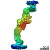
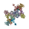
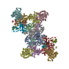
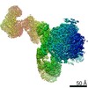
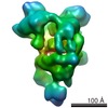
ジャーナル: Proc Natl Acad Sci U S A / 年: 2022 タイトル: Munc13 structural transitions and oligomers that may choreograph successive stages in vesicle priming for neurotransmitter release. 著者: Kirill Grushin / R Venkat Kalyana Sundaram / Charles V Sindelar / James E Rothman / 要旨: How can exactly six SNARE complexes be assembled under each synaptic vesicle? Here we report cryo-EM crystal structures of the core domain of Munc13, the key chaperone that initiates SNAREpin ...How can exactly six SNARE complexes be assembled under each synaptic vesicle? Here we report cryo-EM crystal structures of the core domain of Munc13, the key chaperone that initiates SNAREpin assembly. The functional core of Munc13, consisting of C1-C2B-MUN-C2C (Munc13C) spontaneously crystallizes between phosphatidylserine-rich bilayers in two distinct conformations, each in a radically different oligomeric state. In the open conformation (state 1), Munc13C forms upright trimers that link the two bilayers, separating them by ∼21 nm. In the closed conformation, six copies of Munc13C interact to form a lateral hexamer elevated ∼14 nm above the bilayer. Open and closed conformations differ only by a rigid body rotation around a flexible hinge, which when performed cooperatively assembles Munc13 into a lateral hexamer (state 2) in which the key SNARE assembly-activating site of Munc13 is autoinhibited by its neighbor. We propose that each Munc13 in the lateral hexamer ultimately assembles a single SNAREpin, explaining how only and exactly six SNARE complexes are templated. We suggest that state 1 and state 2 may represent two successive states in the synaptic vesicle supply chain leading to "primed" ready-release vesicles in which SNAREpins are clamped and ready to release (state 3).
電子線照射量: 3.1 e/Å2 / Avg electron dose per subtomogram: 110 e/Å2 / フィルム・検出器のモデル: GATAN K3 (6k x 4k)
電子光学装置
エネルギーフィルター名称: GIF Quantum LS / エネルギーフィルタースリット幅: 20 eV
-
解析
EMソフトウェア
ID
名称
バージョン
カテゴリ
2
SerialEM
画像取得
7
ISOLDE
1.2.0
モデルフィッティング
8
UCSF ChimeraX
1.2
モデルフィッティング
9
UCSF Chimera
1.16
モデルフィッティング
12
RELION
3.1
最終オイラー角割当
14
RELION
3.1
3次元再構成
CTF補正
詳細: CTF correction was performed during 3D reconstruction in RELION 3.1 タイプ: PHASE FLIPPING AND AMPLITUDE CORRECTION
対称性
点対称性: C3 (3回回転対称)
3次元再構成
解像度: 10 Å / 解像度の算出法: FSC 0.143 CUT-OFF / 粒子像の数: 42237 / 対称性のタイプ: POINT
EM volume selection
詳細: Particles were extracted and refined using Warp/M software Num. of tomograms: 62 / Num. of volumes extracted: 70467
原子モデル構築
プロトコル: FLEXIBLE FIT 詳細: Model for fitting was generated by AlphaFold using the construct's amino acid sequence. Flexible fitting into corresponding density was performed using ISOLDE tool in ChimeraX. The resulting ...詳細: Model for fitting was generated by AlphaFold using the construct's amino acid sequence. Flexible fitting into corresponding density was performed using ISOLDE tool in ChimeraX. The resulting structure was copied and fitted as rigid bodies into the 3D map by "fit in map" function in Chimera.
 ムービー
ムービー コントローラー


 データを開く
 基本情報
基本情報 要素
要素 キーワード
キーワード 機能・相同性情報
 データ登録者
 米国, 2件
  引用
引用 構造の表示
構造の表示 ダウンロードとリンク
 その他のダウンロード


 PDBj
PDBj


 集合体
集合体
 Homo sapiens (ヒト) / 参照: UniProt: Q4KUS2
 試料調製
試料調製 電子顕微鏡撮影
 FIELD EMISSION GUN / 加速電圧: 300 kV / 照射モード: FLOOD BEAM
 解析
解析