 Movie
Movie Controller

[English] 日本語
 Yorodumi
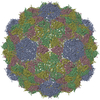
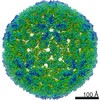
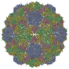
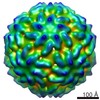
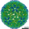
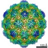
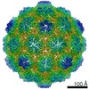
- PDB-7rwz: SaPIbov5 procapsid structure including size redirecting protein Ccm -
+ Open data

- Basic information
| Entry | Database: PDB / ID: 7rwz | |||||||||||||||||||||||||||||||||||||||||||||
|---|---|---|---|---|---|---|---|---|---|---|---|---|---|---|---|---|---|---|---|---|---|---|---|---|---|---|---|---|---|---|---|---|---|---|---|---|---|---|---|---|---|---|---|---|---|---|
| Title | SaPIbov5 procapsid structure including size redirecting protein Ccm | |||||||||||||||||||||||||||||||||||||||||||||
 Components Components |
| |||||||||||||||||||||||||||||||||||||||||||||
 Keywords Keywords | VIRUS / HK97-like fold / capsid size redirection / major capsid protein | |||||||||||||||||||||||||||||||||||||||||||||
| Function / homology |  Function and homology information Function and homology informationATP-dependent peptidase activity / membrane => GO:0016020 / serine-type endopeptidase activity Similarity search - Function | |||||||||||||||||||||||||||||||||||||||||||||
| Biological species |   Staphylococcus aureus (bacteria) Staphylococcus aureus (bacteria) | |||||||||||||||||||||||||||||||||||||||||||||
| Method | ELECTRON MICROSCOPY / single particle reconstruction / cryo EM / Resolution: 4 Å | |||||||||||||||||||||||||||||||||||||||||||||
 Authors Authors | Hawkins, N.C. / Kizziah, J.L. / Dokland, T. | |||||||||||||||||||||||||||||||||||||||||||||
| Funding support |  United States, 2items United States, 2items
| |||||||||||||||||||||||||||||||||||||||||||||
 Citation Citation | Journal: Nat Commun / Year: 2021 Title: Shape shifter: redirection of prolate phage capsid assembly by staphylococcal pathogenicity islands. Authors: N'Toia C Hawkins / James L Kizziah / José R Penadés / Terje Dokland /  Abstract: Staphylococcus aureus pathogenicity islands (SaPIs) are molecular parasites that hijack helper phages for their transfer. SaPIbov5, the prototypical member of a family of cos type SaPIs, redirects ...Staphylococcus aureus pathogenicity islands (SaPIs) are molecular parasites that hijack helper phages for their transfer. SaPIbov5, the prototypical member of a family of cos type SaPIs, redirects the assembly of ϕ12 helper capsids from prolate to isometric. This size and shape shift is dependent on the SaPIbov5-encoded protein Ccm, a homolog of the ϕ12 capsid protein (CP). Using cryo-electron microscopy, we have determined structures of prolate ϕ12 procapsids and isometric SaPIbov5 procapsids. ϕ12 procapsids have icosahedral end caps with T = 4 architecture and a T = 14 cylindrical midsection, whereas SaPIbov5 procapsids have T = 4 icosahedral architecture. We built atomic models for CP and Ccm, and show that Ccm occupies the pentameric capsomers in the isometric SaPIbov5 procapsids, suggesting that preferential incorporation of Ccm pentamers prevents the cylindrical midsection from forming. Our results highlight that pirate elements have evolved diverse mechanisms to suppress phage multiplication, including the acquisition of phage capsid protein homologs. | |||||||||||||||||||||||||||||||||||||||||||||
| History |
|
- Structure visualization
| Movie |
Movie viewer |
|---|---|
| Structure viewer | Molecule: MolmilJmol/JSmol |
- Downloads & links
-Download
| PDBx/mmCIF format | 7rwz.cif.gz | 350.2 KB | Display | PDBx/mmCIF format |
|---|---|---|---|---|
| PDB format | pdb7rwz.ent.gz | 287.7 KB | Display | PDB format |
| PDBx/mmJSON format | 7rwz.json.gz | Tree view | PDBx/mmJSON format | |
| Others |  Other downloads Other downloads |
-Validation report
| Arichive directory | https://data.pdbj.org/pub/pdb/validation_reports/rw/7rwzftp://data.pdbj.org/pub/pdb/validation_reports/rw/7rwz | HTTPS FTP |
|---|
-Related structure data
| Related structure data |  24720MC M: map data used to model this data C: citing same article ( |
|---|---|
| Similar structure data |

-Links
 PDBj
PDBj

- Assembly
| Deposited unit | 
|
|---|---|
| 1 | x 60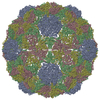
|
| 2 |
|
| 3 | x 5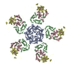
|
| 4 | x 6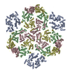
|
| 5 | 
|
| Symmetry | Point symmetry: (Schoenflies symbol: I (icosahedral)) |
-Components
| #1: Protein | Mass: 40468.102 Da / Num. of mol.: 1 Source method: isolated from a genetically manipulated source Source: (gene. exp.) Staphylococcus aureus (bacteria) / Production host: Staphylococcus aureus (bacteria) / References: UniProt: E2FZQ5 | ||
|---|---|---|---|
| #2: Protein | Mass: 45292.820 Da / Num. of mol.: 3 Source method: isolated from a genetically manipulated source Source: (gene. exp.) Staphylococcus aureus (bacteria) / Production host: Staphylococcus aureus (bacteria) / References: UniProt: A0A7H9CBC0Has protein modification | N | |
-Experimental details
-Experiment
| Experiment | Method: ELECTRON MICROSCOPY |
|---|---|
| EM experiment | Aggregation state: PARTICLE / 3D reconstruction method: single particle reconstruction |
- Sample preparation
| Component | Name: SaPIbov5 procapsid / Type: COMPLEX / Entity ID: all / Source: RECOMBINANT |
|---|---|
| Molecular weight | Experimental value: NO |
| Source (natural) | Organism: Staphylococcus aureus (bacteria) |
| Source (recombinant) | Organism: Staphylococcus aureus (bacteria) |
| Buffer solution | pH: 7.8 |
| Specimen | Embedding applied: NO / Shadowing applied: NO / Staining applied: NO / Vitrification applied: YES |
| Vitrification | Cryogen name: ETHANE |
- Electron microscopy imaging
| Experimental equipment |  Model: Titan Krios / Image courtesy: FEI Company |
|---|---|
| Microscopy | Model: FEI TITAN KRIOS |
| Electron gun | Electron source:  FIELD EMISSION GUN / Accelerating voltage: 300 kV / Illumination mode: FLOOD BEAM FIELD EMISSION GUN / Accelerating voltage: 300 kV / Illumination mode: FLOOD BEAM |
| Electron lens | Mode: BRIGHT FIELD |
| Image recording | Electron dose: 60 e/Å2 / Film or detector model: GATAN K3 (6k x 4k) |
- Processing
| Software | Name: PHENIX / Version: 1.14rc1_3161: / Classification: refinement |
|---|---|
| EM software | Name: PHENIX / Category: model refinement |
| CTF correction | Type: PHASE FLIPPING ONLY |
| 3D reconstruction | Resolution: 4 Å / Resolution method: FSC 0.143 CUT-OFF / Num. of particles: 28566 / Symmetry type: POINT |
