Movie
Movie Controller

[English] 日本語
 Yorodumi
- PDB-7rhy: Cre recombinase mutant (D33A/A36V/R192A) in complex with loxA DNA... -
+ Open data

- Basic information
| Entry | Database: PDB / ID: 7rhy | ||||||
|---|---|---|---|---|---|---|---|
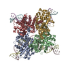
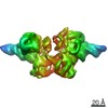
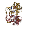
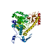
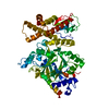
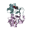
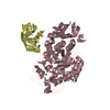
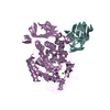
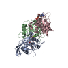
| Title | Cre recombinase mutant (D33A/A36V/R192A) in complex with loxA DNA hairpin | ||||||
 Components Components |
| ||||||
 Keywords Keywords | RECOMBINATION/DNA / Cre / recombinase / monomer / hairpin / RECOMBINATION-DNA complex | ||||||
| Function / homology |  Function and homology information Function and homology information | ||||||
| Biological species |  Escherichia phage P1 (virus) Escherichia phage P1 (virus) | ||||||
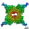
| Method | ELECTRON MICROSCOPY / single particle reconstruction / cryo EM / Resolution: 3.91 Å | ||||||
 Authors Authors | Stachowski, K. / Foster, M.P. | ||||||
| Funding support |  United States, 1items United States, 1items
| ||||||
 Citation Citation | Journal: Nucleic Acids Res / Year: 2022 Title: Mechanisms of Cre recombinase synaptic complex assembly and activation illuminated by Cryo-EM. Authors: Kye Stachowski / Andrew S Norris / Devante Potter / Vicki H Wysocki / Mark P Foster / Abstract: Cre recombinase selectively recognizes DNA and prevents non-specific DNA cleavage through an orchestrated series of assembly intermediates. Cre recombines two loxP DNA sequences featuring a pair of ...Cre recombinase selectively recognizes DNA and prevents non-specific DNA cleavage through an orchestrated series of assembly intermediates. Cre recombines two loxP DNA sequences featuring a pair of palindromic recombinase binding elements and an asymmetric spacer region, by assembly of a tetrameric synaptic complex, cleavage of an opposing pair of strands, and formation of a Holliday junction intermediate. We used Cre and loxP variants to isolate the monomeric Cre-loxP (54 kDa), dimeric Cre2-loxP (110 kDa), and tetrameric Cre4-loxP2 assembly intermediates, and determined their structures using cryo-EM to resolutions of 3.9, 4.5 and 3.2 Å, respectively. Progressive and asymmetric bending of the spacer region along the assembly pathway enables formation of increasingly intimate interfaces between Cre protomers and illuminates the structural bases of biased loxP strand cleavage order and half-the-sites activity. Application of 3D variability analysis to the tetramer data reveals constrained conformational sampling along the pathway between protomer activation and Holliday junction isomerization. These findings underscore the importance of protein and DNA flexibility in Cre-mediated site selection, controlled activation of alternating protomers, the basis for biased strand cleavage order, and recombination efficiency. Such considerations may advance development of site-specific recombinases for use in gene editing applications. | ||||||
| History |
|
- Structure visualization
| Movie |
Movie viewer |
|---|---|
| Structure viewer | Molecule: MolmilJmol/JSmol |
- Downloads & links
-Download
| PDBx/mmCIF format | 7rhy.cif.gz | 148.1 KB | Display | PDBx/mmCIF format |
|---|---|---|---|---|
| PDB format | pdb7rhy.ent.gz | 111.3 KB | Display | PDB format |
| PDBx/mmJSON format | 7rhy.json.gz | Tree view | PDBx/mmJSON format | |
| Others |  Other downloads Other downloads |
-Validation report
| Summary document | 7rhy_validation.pdf.gz | 1.1 MB | Display | wwPDB validaton report |
|---|---|---|---|---|
| Full document | 7rhy_full_validation.pdf.gz | 1.1 MB | Display | |
| Data in XML | 7rhy_validation.xml.gz | 22.5 KB | Display | |
| Data in CIF | 7rhy_validation.cif.gz | 30.1 KB | Display | |
| Arichive directory | https://data.pdbj.org/pub/pdb/validation_reports/rh/7rhyftp://data.pdbj.org/pub/pdb/validation_reports/rh/7rhy | HTTPS FTP |
-Related structure data
| Related structure data | 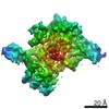 24471MC 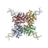 7rhxC  7rhzC M: map data used to model this data C: citing same article ( |
|---|---|
| Similar structure data |


-Links
 PDBj
PDBj


- Assembly
| Deposited unit | 
|
|---|---|
| 1 |
|
-Components
| #1: Protein | Mass: 38493.094 Da / Num. of mol.: 1 / Mutation: D33A, A36V, R192A Source method: isolated from a genetically manipulated source Source: (gene. exp.) Escherichia phage P1 (virus) / Gene: cre / Production host:  |
|---|---|
| #2: DNA chain | Mass: 15119.743 Da / Num. of mol.: 1 / Source method: obtained synthetically / Source: (synth.) Escherichia phage P1 (virus) |
-Experimental details
-Experiment
| Experiment | Method: ELECTRON MICROSCOPY |
|---|---|
| EM experiment | Aggregation state: PARTICLE / 3D reconstruction method: single particle reconstruction |
- Sample preparation
| Component | Name: Complex of Cre recombinase mutant D33A/A36V/R192A and loxA DNA hairpin Type: COMPLEX / Entity ID: all / Source: MULTIPLE SOURCES | |||||||||||||||||||||||||
|---|---|---|---|---|---|---|---|---|---|---|---|---|---|---|---|---|---|---|---|---|---|---|---|---|---|---|
| Molecular weight | Value: 0.054 MDa / Experimental value: NO | |||||||||||||||||||||||||
| Source (natural) | Organism: Escherichia phage P1 (virus) | |||||||||||||||||||||||||
| Buffer solution | pH: 7 Details: Buffer was made fresh from solid reagents and filtered with a 0.22 um filter. | |||||||||||||||||||||||||
| Buffer component |
| |||||||||||||||||||||||||
| Specimen | Conc.: 7.5 mg/ml / Embedding applied: NO / Shadowing applied: NO / Staining applied: NO / Vitrification applied: YES | |||||||||||||||||||||||||
| Specimen support | Details: Pelco easiGLOW at 20 mA / Grid material: GOLD / Grid mesh size: 300 divisions/in. / Grid type: Quantifoil R1.2/1.3 | |||||||||||||||||||||||||
| Vitrification | Instrument: FEI VITROBOT MARK IV / Cryogen name: ETHANE-PROPANE / Humidity: 100 % / Chamber temperature: 277 K |
- Electron microscopy imaging
| Experimental equipment |  Model: Titan Krios / Image courtesy: FEI Company |
|---|---|
| Microscopy | Model: TFS KRIOS |
| Electron gun | Electron source:  FIELD EMISSION GUN / Accelerating voltage: 300 kV / Illumination mode: FLOOD BEAM FIELD EMISSION GUN / Accelerating voltage: 300 kV / Illumination mode: FLOOD BEAM |
| Electron lens | Mode: BRIGHT FIELD / Nominal magnification: 105000 X / Nominal defocus max: 2200 nm / Nominal defocus min: 1000 nm / C2 aperture diameter: 100 µm / Alignment procedure: ZEMLIN TABLEAU |
| Specimen holder | Cryogen: NITROGEN / Specimen holder model: FEI TITAN KRIOS AUTOGRID HOLDER / Temperature (max): 86 K / Temperature (min): 86 K |
| Image recording | Average exposure time: 2 sec. / Electron dose: 60 e/Å2 / Film or detector model: GATAN K3 BIOQUANTUM (6k x 4k) / Num. of grids imaged: 1 / Num. of real images: 6235 / Details: 45 total frames |
| EM imaging optics | Energyfilter name: GIF Bioquantum / Energyfilter slit width: 15 eV Spherical aberration corrector: Microscope was modified with a Cs corrector with two hexapoles. |
| Image scans | Width: 5760 / Height: 4096 |
- Processing
| Software | Name: PHENIX / Version: 1.19.2_4158: / Classification: refinement | ||||||||||||||||||||||||||||||||||||
|---|---|---|---|---|---|---|---|---|---|---|---|---|---|---|---|---|---|---|---|---|---|---|---|---|---|---|---|---|---|---|---|---|---|---|---|---|---|
| EM software |
| ||||||||||||||||||||||||||||||||||||
| CTF correction | Type: PHASE FLIPPING AND AMPLITUDE CORRECTION | ||||||||||||||||||||||||||||||||||||
| Particle selection | Num. of particles selected: 898039 | ||||||||||||||||||||||||||||||||||||
| 3D reconstruction | Resolution: 3.91 Å / Resolution method: FSC 0.143 CUT-OFF / Num. of particles: 146715 / Symmetry type: POINT | ||||||||||||||||||||||||||||||||||||
| Atomic model building | B value: 248.4 / Protocol: FLEXIBLE FIT / Space: REAL / Target criteria: correlation coefficient | ||||||||||||||||||||||||||||||||||||
| Atomic model building | PDB-ID: 2HOI Pdb chain-ID: A / Accession code: 2HOI / Pdb chain residue range: 20-343 / Source name: PDB / Type: experimental model | ||||||||||||||||||||||||||||||||||||
| Refine LS restraints |
|