Movie
Movie Controller

[English] 日本語
 Yorodumi
- PDB-7pbj: Cryo-EM structure of the GroEL-GroES complex with ADP bound to bo... -
+ Open data

- Basic information
| Entry | Database: PDB / ID: 7pbj | ||||||
|---|---|---|---|---|---|---|---|
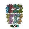
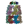
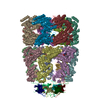
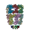
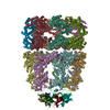
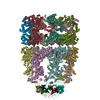
| Title | Cryo-EM structure of the GroEL-GroES complex with ADP bound to both rings ("wide" conformation). | ||||||
 Components Components |
| ||||||
 Keywords Keywords | CHAPERONE / cryo-EM / chaperonin / GroEL / GroEL-GroES | ||||||
| Function / homology |  Function and homology information Function and homology informationGroEL-GroES complex / chaperonin ATPase / mitochondrial unfolded protein response / protein import into mitochondrial intermembrane space / virion assembly / chaperone cofactor-dependent protein refolding / positive regulation of interferon-alpha production / protein folding chaperone / isomerase activity / ATP-dependent protein folding chaperone ...GroEL-GroES complex / chaperonin ATPase / mitochondrial unfolded protein response / protein import into mitochondrial intermembrane space / virion assembly / chaperone cofactor-dependent protein refolding / positive regulation of interferon-alpha production / protein folding chaperone / isomerase activity / ATP-dependent protein folding chaperone / response to radiation / positive regulation of interleukin-6 production / positive regulation of type II interferon production / unfolded protein binding / protein folding / positive regulation of T cell activation / protein-folding chaperone binding / response to heat / protein refolding / magnesium ion binding / ATP hydrolysis activity / ATP binding / identical protein binding / membrane / metal ion binding / cytosol Similarity search - Function | ||||||
| Biological species |  | ||||||
| Method | ELECTRON MICROSCOPY / single particle reconstruction / cryo EM / Resolution: 3.4 Å | ||||||
 Authors Authors | Pichkur, E.B. / Stanishneva-Konovalova, T.B. | ||||||
| Funding support |  Russian Federation, 1items Russian Federation, 1items
| ||||||
 Citation Citation | Journal: Sci Rep / Year: 2021 Title: Novel cryo-EM structure of an ADP-bound GroEL-GroES complex. Authors: Sofia S Kudryavtseva / Evgeny B Pichkur / Igor A Yaroshevich / Aleksandra A Mamchur / Irina S Panina / Andrei V Moiseenko / Olga S Sokolova / Vladimir I Muronetz / Tatiana B Stanishneva-Konovalova / Abstract: The GroEL-GroES chaperonin complex is a bacterial protein folding system, functioning in an ATP-dependent manner. Upon ATP binding and hydrolysis, it undergoes multiple stages linked to substrate ...The GroEL-GroES chaperonin complex is a bacterial protein folding system, functioning in an ATP-dependent manner. Upon ATP binding and hydrolysis, it undergoes multiple stages linked to substrate protein binding, folding and release. Structural methods helped to reveal several conformational states and provide more information about the chaperonin functional cycle. Here, using cryo-EM we resolved two nucleotide-bound structures of the bullet-shaped GroEL-GroES complex at 3.4 Å resolution. The main difference between them is the relative orientation of their apical domains. Both structures contain nucleotides in cis and trans GroEL rings; in contrast to previously reported bullet-shaped complexes where nucleotides were only present in the cis ring. Our results suggest that the bound nucleotides correspond to ADP, and that such a state appears at low ATP:ADP ratios. | ||||||
| History |
|
- Structure visualization
| Movie |
Movie viewer |
|---|---|
| Structure viewer | Molecule: MolmilJmol/JSmol |
- Downloads & links
-Download
| PDBx/mmCIF format | 7pbj.cif.gz | 1.2 MB | Display | PDBx/mmCIF format |
|---|---|---|---|---|
| PDB format | pdb7pbj.ent.gz | Display | PDB format | |
| PDBx/mmJSON format | 7pbj.json.gz | Tree view | PDBx/mmJSON format | |
| Others |  Other downloads Other downloads |
-Validation report
| Summary document | 7pbj_validation.pdf.gz | 1.9 MB | Display | wwPDB validaton report |
|---|---|---|---|---|
| Full document | 7pbj_full_validation.pdf.gz | 2 MB | Display | |
| Data in XML | 7pbj_validation.xml.gz | 200 KB | Display | |
| Data in CIF | 7pbj_validation.cif.gz | 301.4 KB | Display | |
| Arichive directory | https://data.pdbj.org/pub/pdb/validation_reports/pb/7pbjftp://data.pdbj.org/pub/pdb/validation_reports/pb/7pbj | HTTPS FTP |
-Related structure data
| Related structure data |  13293MC  7pbxC M: map data used to model this data C: citing same article ( |
|---|---|
| Similar structure data |


-Links
 PDBj
PDBj

- Assembly
| Deposited unit | 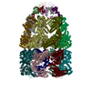
|
|---|---|
| 1 |
|
-Components
| #1: Protein | Mass: 55220.105 Da / Num. of mol.: 14 Source method: isolated from a genetically manipulated source Details: GroEL from Escherichia coli str. K-12 substr. W3110 Source: (gene. exp.) Strain: K12 / Gene: groL, groEL, mopA, b4143, JW4103 Production host: References: UniProt: P0A6F5 #2: Protein | Mass: 10400.938 Da / Num. of mol.: 7 Source method: isolated from a genetically manipulated source Details: GroES from Escherichia coli str. K-12 substr. W3110 Source: (gene. exp.) Strain: K12 / Gene: groS, groES, mopB, b4142, JW4102 Production host: References: UniProt: P0A6F9 #3: Chemical | ChemComp-ADP /   Mass: 427.201 Da / Num. of mol.: 14 / Source method: obtained synthetically / Formula: C10H15N5O10P2 / Feature type: SUBJECT OF INVESTIGATION / Comment: ADP, energy-carrying molecule*YM Mass: 427.201 Da / Num. of mol.: 14 / Source method: obtained synthetically / Formula: C10H15N5O10P2 / Feature type: SUBJECT OF INVESTIGATION / Comment: ADP, energy-carrying molecule*YM#4: Chemical | ChemComp-MG /   Mass: 24.305 Da / Num. of mol.: 14 / Source method: obtained synthetically / Formula: Mg Mass: 24.305 Da / Num. of mol.: 14 / Source method: obtained synthetically / Formula: Mg#5: Water | ChemComp-HOH / |  Mass: 18.015 Da / Num. of mol.: 14 / Source method: isolated from a natural source / Formula: H2O Mass: 18.015 Da / Num. of mol.: 14 / Source method: isolated from a natural source / Formula: H2OHas ligand of interest | Y | |
|---|
-Experimental details
-Experiment
| Experiment | Method: ELECTRON MICROSCOPY |
|---|---|
| EM experiment | Aggregation state: PARTICLE / 3D reconstruction method: single particle reconstruction |
- Sample preparation
| Component | Name: ADP-bound GroEL-GroES complex / Type: COMPLEX / Entity ID: #1-#2 / Source: RECOMBINANT |
|---|---|
| Molecular weight | Value: 0.882 MDa / Experimental value: NO |
| Source (natural) | Organism: |
| Source (recombinant) | Organism: |
| Buffer solution | pH: 7.5 |
| Specimen | Conc.: 1 mg/ml / Embedding applied: NO / Shadowing applied: NO / Staining applied: NO / Vitrification applied: YES |
| Specimen support | Grid material: COPPER / Grid mesh size: 300 divisions/in. / Grid type: Quantifoil R1.2/1.3 |
| Vitrification | Instrument: FEI VITROBOT MARK IV / Cryogen name: ETHANE / Humidity: 100 % / Chamber temperature: 4.5 K |
- Electron microscopy imaging
| Experimental equipment |  Model: Titan Krios / Image courtesy: FEI Company |
|---|---|
| Microscopy | Model: FEI TITAN KRIOS |
| Electron gun | Electron source:  FIELD EMISSION GUN / Accelerating voltage: 300 kV / Illumination mode: SPOT SCAN FIELD EMISSION GUN / Accelerating voltage: 300 kV / Illumination mode: SPOT SCAN |
| Electron lens | Mode: BRIGHT FIELD / Cs: 0.01 mm / C2 aperture diameter: 100 µm |
| Image recording | Electron dose: 100 e/Å2 / Detector mode: INTEGRATING / Film or detector model: FEI FALCON II (4k x 4k) |
- Processing
| EM software |
| ||||||||||||||||||||
|---|---|---|---|---|---|---|---|---|---|---|---|---|---|---|---|---|---|---|---|---|---|
| CTF correction | Type: PHASE FLIPPING AND AMPLITUDE CORRECTION | ||||||||||||||||||||
| Symmetry | Point symmetry: C7 (7 fold cyclic) | ||||||||||||||||||||
| 3D reconstruction | Resolution: 3.4 Å / Resolution method: FSC 0.143 CUT-OFF / Num. of particles: 41000 / Symmetry type: POINT | ||||||||||||||||||||
| Atomic model building | PDB-ID: 1SX4 Accession code: 1SX4 / Source name: PDB / Type: experimental model |