 Movie
Movie Controller


+ Open data

- Basic information
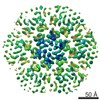
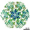
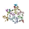
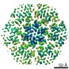
| Entry | Database: PDB / ID: 7ovq | |||||||||||||||||||||||||||
|---|---|---|---|---|---|---|---|---|---|---|---|---|---|---|---|---|---|---|---|---|---|---|---|---|---|---|---|---|
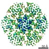
| Title | Immature HIV-1 matrix structure | |||||||||||||||||||||||||||
 Components Components | Gag polyprotein | |||||||||||||||||||||||||||
 Keywords Keywords | VIRUS / HIV-1 / Gag / matrix | |||||||||||||||||||||||||||
| Function / homology |  Function and homology information Function and homology informationhost multivesicular body / viral nucleocapsid / viral translational frameshifting / host cell nucleus / host cell plasma membrane / virion membrane / structural molecule activity / RNA binding / zinc ion binding / ATP binding Similarity search - Function | |||||||||||||||||||||||||||
| Biological species |   Human immunodeficiency virus 1 Human immunodeficiency virus 1 | |||||||||||||||||||||||||||
| Method | ELECTRON MICROSCOPY / subtomogram averaging / cryo EM / Resolution: 7.2 Å | |||||||||||||||||||||||||||
 Authors Authors | Qu, K. / Ke, Z.L. / Zila, V. / Anders-Oesswein, M. / Glass, B. / Muecksch, F. / Mueller, R. / Schultz, C. / Mueller, B. / Kraeusslich, H.G. / Briggs, J.A.G. | |||||||||||||||||||||||||||
| Funding support |  Germany, 8items Germany, 8items
| |||||||||||||||||||||||||||
 Citation Citation | Journal: Science / Year: 2021 Title: Maturation of the matrix and viral membrane of HIV-1. Authors: Kun Qu / Zunlong Ke / Vojtech Zila / Maria Anders-Össwein / Bärbel Glass / Frauke Mücksch / Rainer Müller / Carsten Schultz / Barbara Müller / Hans-Georg Kräusslich / John A G Briggs /   Abstract: Gag, the primary structural protein of HIV-1, is recruited to the plasma membrane for virus assembly by its matrix (MA) domain. Gag is subsequently cleaved into its component domains, causing ...Gag, the primary structural protein of HIV-1, is recruited to the plasma membrane for virus assembly by its matrix (MA) domain. Gag is subsequently cleaved into its component domains, causing structural maturation to repurpose the virion for cell entry. We determined the structure and arrangement of MA within immature and mature HIV-1 through cryo-electron tomography. We found that MA rearranges between two different hexameric lattices upon maturation. In mature HIV-1, a lipid extends out of the membrane to bind with a pocket in MA. Our data suggest that proteolytic maturation of HIV-1 not only assembles the viral capsid surrounding the genome but also repurposes the membrane-bound MA lattice for an entry or postentry function and results in the partial removal of up to 2500 lipids from the viral membrane. | |||||||||||||||||||||||||||
| History |
|
- Structure visualization
| Movie |
Movie viewer |
|---|---|
| Structure viewer | Molecule: MolmilJmol/JSmol |
- Downloads & links
-Download
| PDBx/mmCIF format | 7ovq.cif.gz | 364.6 KB | Display | PDBx/mmCIF format |
|---|---|---|---|---|
| PDB format | pdb7ovq.ent.gz | 238.1 KB | Display | PDB format |
| PDBx/mmJSON format | 7ovq.json.gz | Tree view | PDBx/mmJSON format | |
| Others |  Other downloads Other downloads |
-Validation report
| Arichive directory | https://data.pdbj.org/pub/pdb/validation_reports/ov/7ovqftp://data.pdbj.org/pub/pdb/validation_reports/ov/7ovq | HTTPS FTP |
|---|
-Related structure data
| Related structure data |  13087MC  7ovrC M: map data used to model this data C: citing same article ( |
|---|---|
| Similar structure data |

-Links
 PDBj
PDBj


- Assembly
| Deposited unit | 
|
|---|---|
| 1 |
|
-Components
| #1: Protein | Mass: 13084.983 Da / Num. of mol.: 24 Source method: isolated from a genetically manipulated source Source: (gene. exp.) Human immunodeficiency virus 1 / Gene: gag / Production host:  Homo sapiens (human) / References: UniProt: B6DRA0 Homo sapiens (human) / References: UniProt: B6DRA0#2: Chemical | ChemComp-MYR /   Mass: 228.371 Da / Num. of mol.: 24 / Source method: obtained synthetically / Formula: C14H28O2 / Feature type: SUBJECT OF INVESTIGATION Mass: 228.371 Da / Num. of mol.: 24 / Source method: obtained synthetically / Formula: C14H28O2 / Feature type: SUBJECT OF INVESTIGATIONHas ligand of interest | Y | Has protein modification | Y | |
|---|
-Experimental details
-Experiment
| Experiment | Method: ELECTRON MICROSCOPY |
|---|---|
| EM experiment | Aggregation state: PARTICLE / 3D reconstruction method: subtomogram averaging |
- Sample preparation
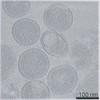
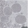
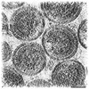
| Component | Name: Human immunodeficiency virus 1 / Type: VIRUS Details: HIV-1 NL4-3 protease defective (D25A) virus particles purified from HEK293T cells. Entity ID: #1 / Source: RECOMBINANT |
|---|---|
| Source (natural) | Organism: Human immunodeficiency virus 1 |
| Source (recombinant) | Organism: Homo sapiens (human) |
| Details of virus | Empty: NO / Enveloped: YES / Isolate: STRAIN / Type: VIRION |
| Buffer solution | pH: 7.4 |
| Specimen | Embedding applied: NO / Shadowing applied: NO / Staining applied: NO / Vitrification applied: YES Details: HIV-1 NL4-3 protease defective (D25A) virus particles purified from HEK293T cells. |
| Vitrification | Cryogen name: ETHANE |
- Electron microscopy imaging
| Experimental equipment |  Model: Titan Krios / Image courtesy: FEI Company |
|---|---|
| Microscopy | Model: FEI TITAN KRIOS |
| Electron gun | Electron source:  FIELD EMISSION GUN / Accelerating voltage: 300 kV / Illumination mode: FLOOD BEAM FIELD EMISSION GUN / Accelerating voltage: 300 kV / Illumination mode: FLOOD BEAM |
| Electron lens | Mode: BRIGHT FIELD / Nominal magnification: 105000 X / Nominal defocus max: 5000 nm / Nominal defocus min: 1500 nm / Cs: 2.7 mm / C2 aperture diameter: 50 µm |
| Specimen holder | Cryogen: NITROGEN / Specimen holder model: FEI TITAN KRIOS AUTOGRID HOLDER |
| Image recording | Electron dose: 3 e/Å2 / Detector mode: SUPER-RESOLUTION / Film or detector model: GATAN K2 QUANTUM (4k x 4k) / Details: Number of frames ranged from 10-12. |
| EM imaging optics | Energyfilter name: GIF Quantum LS / Energyfilter slit width: 20 eV |
- Processing
| EM software |
| |||||||||||||||||||||
|---|---|---|---|---|---|---|---|---|---|---|---|---|---|---|---|---|---|---|---|---|---|---|
| CTF correction | Type: PHASE FLIPPING AND AMPLITUDE CORRECTION | |||||||||||||||||||||
| Symmetry | Point symmetry: C3 (3 fold cyclic) | |||||||||||||||||||||
| 3D reconstruction | Resolution: 7.2 Å / Resolution method: FSC 0.143 CUT-OFF / Num. of particles: 22417 / Symmetry type: POINT | |||||||||||||||||||||
| EM volume selection | Num. of tomograms: 74 / Num. of volumes extracted: 251207 | |||||||||||||||||||||
| Atomic model building | Protocol: RIGID BODY FIT Details: Only the backbone of the protein model was fitted as a rigid body. | |||||||||||||||||||||
| Atomic model building | PDB-ID: 2H3Q Accession code: 2H3Q / Source name: PDB / Type: experimental model |

