 Movie
Movie Controller

[English] 日本語
 Yorodumi
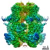
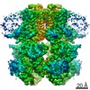
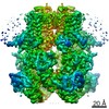
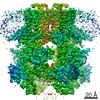
- PDB-7mbp: Cryo-EM structure of zebrafish TRPM5 in the presence of 1 mM EDTA -
+ Open data

- Basic information
| Entry | Database: PDB / ID: 7mbp | |||||||||||||||||||||||||||
|---|---|---|---|---|---|---|---|---|---|---|---|---|---|---|---|---|---|---|---|---|---|---|---|---|---|---|---|---|
| Title | Cryo-EM structure of zebrafish TRPM5 in the presence of 1 mM EDTA | |||||||||||||||||||||||||||
 Components Components | Transient receptor potential melastatin 5 | |||||||||||||||||||||||||||
 Keywords Keywords | TRANSPORT PROTEIN / Ion channel / TRP channel | |||||||||||||||||||||||||||
| Function / homology |  Function and homology information Function and homology informationcalcium-activated cation channel activity / calcium channel activity / calcium ion binding / identical protein binding / plasma membrane Similarity search - Function | |||||||||||||||||||||||||||
| Biological species |  | |||||||||||||||||||||||||||
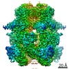
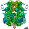
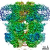
| Method | ELECTRON MICROSCOPY / single particle reconstruction / cryo EM / Resolution: 2.8 Å | |||||||||||||||||||||||||||
 Authors Authors | Ruan, Z. / Lu, W. / Du, J. / Haley, E. | |||||||||||||||||||||||||||
| Funding support |  United States, 5items United States, 5items
| |||||||||||||||||||||||||||
 Citation Citation | Journal: Nat Struct Mol Biol / Year: 2021 Title: Structures of the TRPM5 channel elucidate mechanisms of activation and inhibition. Authors: Zheng Ruan / Emery Haley / Ian J Orozco / Mark Sabat / Richard Myers / Rebecca Roth / Juan Du / Wei Lü / Abstract: The Ca-activated TRPM5 channel plays essential roles in taste perception and insulin secretion. However, the mechanism by which Ca regulates TRPM5 activity remains elusive. We report cryo-EM ...The Ca-activated TRPM5 channel plays essential roles in taste perception and insulin secretion. However, the mechanism by which Ca regulates TRPM5 activity remains elusive. We report cryo-EM structures of the zebrafish TRPM5 in an apo closed state, a Ca-bound open state, and an antagonist-bound inhibited state. We define two novel ligand binding sites: a Ca site (Ca) in the intracellular domain and an antagonist site in the transmembrane domain (TMD). The Ca site is unique to TRPM5 and has two roles: modulating the voltage dependence and promoting Ca binding to the Ca site, which is conserved throughout TRPM channels. Conformational changes initialized from both Ca sites cooperatively open the ion-conducting pore. The antagonist NDNA wedges into the space between the S1-S4 domain and pore domain, stabilizing the transmembrane domain in an apo-like closed state. Our results lay the foundation for understanding the voltage-dependent TRPM channels and developing new therapeutic agents. | |||||||||||||||||||||||||||
| History |
|
- Structure visualization
| Movie |
Movie viewer |
|---|---|
| Structure viewer | Molecule: MolmilJmol/JSmol |
- Downloads & links
-Download
| PDBx/mmCIF format | 7mbp.cif.gz | 690.6 KB | Display | PDBx/mmCIF format |
|---|---|---|---|---|
| PDB format | pdb7mbp.ent.gz | 554.7 KB | Display | PDB format |
| PDBx/mmJSON format | 7mbp.json.gz | Tree view | PDBx/mmJSON format | |
| Others |  Other downloads Other downloads |
-Validation report
| Arichive directory | https://data.pdbj.org/pub/pdb/validation_reports/mb/7mbpftp://data.pdbj.org/pub/pdb/validation_reports/mb/7mbp | HTTPS FTP |
|---|
-Related structure data
| Related structure data | 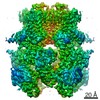 23740MC  7mbqC  7mbrC  7mbsC  7mbtC 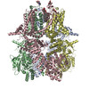 7mbuC 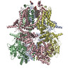 7mbvC M: map data used to model this data C: citing same article ( |
|---|---|
| Similar structure data |


-Links
 PDBj
PDBj
- Assembly
| Deposited unit | 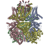
|
|---|---|
| 1 |
|
-Components
| #1: Protein | Mass: 132799.000 Da / Num. of mol.: 4 Source method: isolated from a genetically manipulated source Source: (gene. exp.)  Homo sapiens (human) / References: UniProt: S5UH55 Homo sapiens (human) / References: UniProt: S5UH55#2: Sugar | ChemComp-NAG /   Type: D-saccharide, beta linking / Mass: 221.208 Da / Num. of mol.: 4 / Source method: obtained synthetically / Formula: C8H15NO6 / Feature type: SUBJECT OF INVESTIGATION Type: D-saccharide, beta linking / Mass: 221.208 Da / Num. of mol.: 4 / Source method: obtained synthetically / Formula: C8H15NO6 / Feature type: SUBJECT OF INVESTIGATION#3: Chemical | ChemComp-YUV / ( 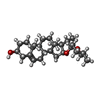  Mass: 414.621 Da / Num. of mol.: 4 / Source method: obtained synthetically / Formula: C27H42O3 / Feature type: SUBJECT OF INVESTIGATION Mass: 414.621 Da / Num. of mol.: 4 / Source method: obtained synthetically / Formula: C27H42O3 / Feature type: SUBJECT OF INVESTIGATION#4: Chemical | ChemComp-YUY / (   Mass: 841.033 Da / Num. of mol.: 4 / Source method: obtained synthetically / Formula: C44H72O15 / Feature type: SUBJECT OF INVESTIGATION Mass: 841.033 Da / Num. of mol.: 4 / Source method: obtained synthetically / Formula: C44H72O15 / Feature type: SUBJECT OF INVESTIGATIONHas ligand of interest | Y | Has protein modification | Y | |
|---|
-Experimental details
-Experiment
| Experiment | Method: ELECTRON MICROSCOPY |
|---|---|
| EM experiment | Aggregation state: PARTICLE / 3D reconstruction method: single particle reconstruction |
- Sample preparation
| Component | Name: TRPM5 channel / Type: COMPLEX / Entity ID: #1 / Source: RECOMBINANT |
|---|---|
| Source (natural) | Organism: |
| Source (recombinant) | Organism: Homo sapiens (human) |
| Buffer solution | pH: 8 |
| Specimen | Embedding applied: NO / Shadowing applied: NO / Staining applied: NO / Vitrification applied: YES |
| Vitrification | Cryogen name: ETHANE |
- Electron microscopy imaging
| Experimental equipment |  Model: Titan Krios / Image courtesy: FEI Company |
|---|---|
| Microscopy | Model: FEI TITAN KRIOS |
| Electron gun | Electron source:  FIELD EMISSION GUN / Accelerating voltage: 300 kV / Illumination mode: FLOOD BEAM FIELD EMISSION GUN / Accelerating voltage: 300 kV / Illumination mode: FLOOD BEAM |
| Electron lens | Mode: BRIGHT FIELD |
| Image recording | Electron dose: 49.6 e/Å2 / Detector mode: SUPER-RESOLUTION / Film or detector model: GATAN K2 SUMMIT (4k x 4k) |
- Processing
| Software | Name: PHENIX / Version: 1.19rc3_4028: / Classification: refinement | ||||||||||||||||||||||||
|---|---|---|---|---|---|---|---|---|---|---|---|---|---|---|---|---|---|---|---|---|---|---|---|---|---|
| EM software | Name: PHENIX / Category: model refinement | ||||||||||||||||||||||||
| CTF correction | Type: PHASE FLIPPING ONLY | ||||||||||||||||||||||||
| 3D reconstruction | Resolution: 2.8 Å / Num. of particles: 84000 / Symmetry type: POINT | ||||||||||||||||||||||||
| Atomic model building | Protocol: AB INITIO MODEL | ||||||||||||||||||||||||
| Refine LS restraints |
|
