 Movie
Movie Controller

[English] 日本語
 Yorodumi
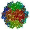
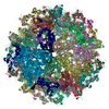
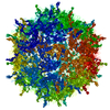
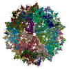
- PDB-7kfr: Adeno-Associated Virus (AAV-DJ) - cryo-EM structure at 1.56 Angst... -
+ Open data

- Basic information
| Entry | Database: PDB / ID: 7kfr | ||||||
|---|---|---|---|---|---|---|---|
| Title | Adeno-Associated Virus (AAV-DJ) - cryo-EM structure at 1.56 Angstrom Resolution | ||||||
 Components Components | Capsid protein VP1 | ||||||
 Keywords Keywords | VIRUS / Gene therapy vector / VIRUS LIKE PARTICLE | ||||||
| Function / homology | Phospholipase A2-like domain / Phospholipase A2-like domain / Parvovirus coat protein VP2 / Parvovirus coat protein VP1/VP2 / Parvovirus coat protein VP2 / Capsid/spike protein, ssDNA virus / T=1 icosahedral viral capsid / structural molecule activity / Capsid protein VP1 Function and homology information Function and homology information | ||||||
| Biological species |   Adeno-associated virus Adeno-associated virus | ||||||
| Method | ELECTRON MICROSCOPY / single particle reconstruction / cryo EM / Resolution: 1.56 Å | ||||||
 Authors Authors | Xie, Q. / Yoshioka, C.K. / Chapman, M.S. | ||||||
| Funding support |  United States, 1items United States, 1items
| ||||||
 Citation Citation | Journal: Viruses / Year: 2020 Title: Adeno-Associated Virus (AAV-DJ)-Cryo-EM Structure at 1.56 Å Resolution. Authors: Qing Xie / Craig K Yoshioka / Michael S Chapman / Abstract: Adeno-associated virus is the leading viral vector for gene therapy. AAV-DJ is a recombinant variant developed for tropism to the liver. The AAV-DJ structure has been determined to 1.56 Å resolution ...Adeno-associated virus is the leading viral vector for gene therapy. AAV-DJ is a recombinant variant developed for tropism to the liver. The AAV-DJ structure has been determined to 1.56 Å resolution through cryo-electron microscopy (cryo-EM). Only apoferritin is reported in preprints at 1.6 Å or higher resolution, and AAV-DJ nearly matches the highest resolutions ever attained through X-ray diffraction of virus crystals. However, cryo-EM has the advantage that most of the hydrogens are clear, improving the accuracy of atomic refinement, and removing ambiguity in hydrogen bond identification. Outside of secondary structures where hydrogen bonding was predictable a priori, the networks of hydrogen bonds coming from direct observation of hydrogens and acceptor atoms are quite different from those inferred even at 2.8 Å resolution. The implications for understanding viral assembly mean that cryo-EM will likely become the favored approach for high resolution structural virology. | ||||||
| History |
|
- Structure visualization
| Movie |
Movie viewer |
|---|---|
| Structure viewer | Molecule: MolmilJmol/JSmol |
- Downloads & links
-Download
| PDBx/mmCIF format | 7kfr.cif.gz | 214.9 KB | Display | PDBx/mmCIF format |
|---|---|---|---|---|
| PDB format | pdb7kfr.ent.gz | 171 KB | Display | PDB format |
| PDBx/mmJSON format | 7kfr.json.gz | Tree view | PDBx/mmJSON format | |
| Others |  Other downloads Other downloads |
-Validation report
| Summary document | 7kfr_validation.pdf.gz | 1.7 MB | Display | wwPDB validaton report |
|---|---|---|---|---|
| Full document | 7kfr_full_validation.pdf.gz | 1.7 MB | Display | |
| Data in XML | 7kfr_validation.xml.gz | 35.2 KB | Display | |
| Data in CIF | 7kfr_validation.cif.gz | 49.8 KB | Display | |
| Arichive directory | https://data.pdbj.org/pub/pdb/validation_reports/kf/7kfrftp://data.pdbj.org/pub/pdb/validation_reports/kf/7kfr | HTTPS FTP |
-Related structure data
| Related structure data | 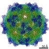 22854MC M: map data used to model this data C: citing same article ( |
|---|---|
| Similar structure data | |
| EM raw data | EMPIAR-10551 (Title: Adeno-Associated Virus (AAV-DJ)-Cryo-EM Structure at 1.56 Å Resolution Data size: 2.8 TB Data #1: Falcon3 frames converted to TIFF and compressed [micrographs - multiframe]) |


-Links
 PDBj
PDBj


- Assembly
| Deposited unit | 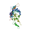
|
|---|---|
| 1 | x 60
|
| 2 |
|
| 3 | x 5
|
| 4 | x 6
|
| 5 | 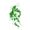
|
| Symmetry | Point symmetry: (Schoenflies symbol: I (icosahedral)) |
-Components
| #1: Protein | Mass: 58877.809 Da / Num. of mol.: 1 Source method: isolated from a genetically manipulated source Source: (gene. exp.) Adeno-associated virus / Gene: cap / Variant: DJ / Production host:   Spodoptera frugiperda (fall armyworm) / References: UniProt: Q6JC41 Spodoptera frugiperda (fall armyworm) / References: UniProt: Q6JC41 | ||||
|---|---|---|---|---|---|
| #2: Chemical |   Mass: 24.305 Da / Num. of mol.: 3 / Source method: obtained synthetically / Formula: Mg Mass: 24.305 Da / Num. of mol.: 3 / Source method: obtained synthetically / Formula: Mg#3: Water | ChemComp-HOH / |  Mass: 18.015 Da / Num. of mol.: 265 / Source method: isolated from a natural source / Formula: H2O Mass: 18.015 Da / Num. of mol.: 265 / Source method: isolated from a natural source / Formula: H2OHas ligand of interest | N | |
-Experimental details
-Experiment
| Experiment | Method: ELECTRON MICROSCOPY |
|---|---|
| EM experiment | Aggregation state: PARTICLE / 3D reconstruction method: single particle reconstruction |
- Sample preparation
| Component | Name: Adeno-associated virus / Type: VIRUS / Entity ID: #1 / Source: RECOMBINANT | ||||||||||||||||||||
|---|---|---|---|---|---|---|---|---|---|---|---|---|---|---|---|---|---|---|---|---|---|
| Molecular weight | Value: 3.746 MDa / Experimental value: NO | ||||||||||||||||||||
| Source (natural) | Organism: Adeno-associated virus | ||||||||||||||||||||
| Source (recombinant) | Organism: Spodoptera frugiperda (fall armyworm) | ||||||||||||||||||||
| Details of virus | Empty: YES / Enveloped: NO / Isolate: STRAIN / Type: VIRUS-LIKE PARTICLE | ||||||||||||||||||||
| Natural host | Organism: Homo sapiens | ||||||||||||||||||||
| Virus shell | Diameter: 250 nm / Triangulation number (T number): 1 | ||||||||||||||||||||
| Buffer solution | pH: 7.4 | ||||||||||||||||||||
| Buffer component |
| ||||||||||||||||||||
| Specimen | Conc.: 0.6 mg/ml / Embedding applied: NO / Shadowing applied: NO / Staining applied: NO / Vitrification applied: YES / Details: Monodisperse | ||||||||||||||||||||
| Specimen support | Details: PELCO easiGlow Glow Discharge Cleaning System Current: 15mA Grid material: COPPER / Grid mesh size: 200 divisions/in. / Grid type: Quantifoil | ||||||||||||||||||||
| Vitrification | Instrument: FEI VITROBOT MARK IV / Cryogen name: ETHANE / Humidity: 100 % / Chamber temperature: 293 K Details: Two 3uL aliquots applied to grid (manual blotting between), prior to automated 3 second blot before plunging. |
- Electron microscopy imaging
| Experimental equipment |  Model: Titan Krios / Image courtesy: FEI Company |
|---|---|
| Microscopy | Model: FEI TITAN KRIOS Details: Coma-free alignment and objective astigmatism where corrected using Sherpa (Thermo Fisher, Inc.). |
| Electron gun | Electron source:  FIELD EMISSION GUN / Accelerating voltage: 300 kV / Illumination mode: FLOOD BEAM FIELD EMISSION GUN / Accelerating voltage: 300 kV / Illumination mode: FLOOD BEAM |
| Electron lens | Mode: BRIGHT FIELD / Nominal magnification: 155000 X / Calibrated magnification: 154400 X / Nominal defocus max: -2600 nm / Nominal defocus min: -800 nm / Cs: 2.7 mm / Alignment procedure: COMA FREE |
| Specimen holder | Cryogen: NITROGEN / Specimen holder model: FEI TITAN KRIOS AUTOGRID HOLDER / Temperature (max): 93 K / Temperature (min): 93 K |
| Image recording | Average exposure time: 15.8 sec. / Electron dose: 30 e/Å2 / Detector mode: INTEGRATING / Film or detector model: FEI FALCON III (4k x 4k) / Num. of grids imaged: 1 / Num. of real images: 2241 Details: Data were collected with pixel size of 0.514 angstrom on a FEI Titan Krios (Thermo Fisher, Inc.) at 300 kV, using a Falcon 3 camera (Thermo Fisher) with a total dose of approx. 30 e-/A2 ...Details: Data were collected with pixel size of 0.514 angstrom on a FEI Titan Krios (Thermo Fisher, Inc.) at 300 kV, using a Falcon 3 camera (Thermo Fisher) with a total dose of approx. 30 e-/A2 fractionated across 200 frames. Camera dose rate was approx. 0.5 e-/pixel/s. Defocus was random in the nominal range of -0.8 to -2.6 um. Images were acquired in EPU (Thermo Fisher, Inc.) without using image shift. |
| Image scans | Sampling size: 14 µm / Width: 4096 / Height: 4096 |
- Processing
| EM software |
| |||||||||||||||||||||||||||||||||||||||||||||||||||||||
|---|---|---|---|---|---|---|---|---|---|---|---|---|---|---|---|---|---|---|---|---|---|---|---|---|---|---|---|---|---|---|---|---|---|---|---|---|---|---|---|---|---|---|---|---|---|---|---|---|---|---|---|---|---|---|---|---|
| CTF correction | Type: PHASE FLIPPING ONLY | |||||||||||||||||||||||||||||||||||||||||||||||||||||||
| Particle selection | Num. of particles selected: 75316 Details: DoG (Difference of Gaussian) Picker was used for initial automated particle selection. Templates were then generated by 2D classification, followed by particle template selection in Relion 3.0. | |||||||||||||||||||||||||||||||||||||||||||||||||||||||
| Symmetry | Point symmetry: I (icosahedral) | |||||||||||||||||||||||||||||||||||||||||||||||||||||||
| 3D reconstruction | Resolution: 1.56 Å / Resolution method: FSC 0.143 CUT-OFF / Num. of particles: 48209 Details: These 48209 particles refined to approx. 2.2 A with I1 symmetry, and subsequent refinements of beam tilt and per-particle CTF brought the resolution to 1.8 A. Further particle-polishing and ...Details: These 48209 particles refined to approx. 2.2 A with I1 symmetry, and subsequent refinements of beam tilt and per-particle CTF brought the resolution to 1.8 A. Further particle-polishing and subsequent re-refinement of CTF brought the resolution to 1.70 A and a final reconstruction using Ewald sphere correction ended at 1.56 A. The map used for modeling was sharpened using the volume whitening routine in cisTEM. Num. of class averages: 1 / Symmetry type: POINT | |||||||||||||||||||||||||||||||||||||||||||||||||||||||
| Atomic model building | Protocol: FLEXIBLE FIT / Space: REAL / Target criteria: Least-squares residual Details: Stand-alone RSRef was used for refinement of magnification, resolution, envelope correction and atomic B-factors. This was alternated with RSRef-embedded CNS was used for molecular dynamics ...Details: Stand-alone RSRef was used for refinement of magnification, resolution, envelope correction and atomic B-factors. This was alternated with RSRef-embedded CNS was used for molecular dynamics optimization (1st round) and stereochemically-restrained all-atom least-squares optimization. | |||||||||||||||||||||||||||||||||||||||||||||||||||||||
| Atomic model building | PDB-ID: 5UF6 Accession code: 5UF6 / Source name: PDB / Type: experimental model |

