 Movie
Movie Controller

[English] 日本語
 Yorodumi
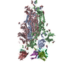
- PDB-7k7k: Structure of the EPEC type III secretion injectisome EspA filament -
+ Open data

- Basic information
| Entry | Database: PDB / ID: 7k7k | |||||||||
|---|---|---|---|---|---|---|---|---|---|---|
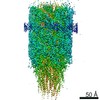
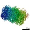
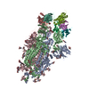
| Title | Structure of the EPEC type III secretion injectisome EspA filament | |||||||||
 Components Components | Translocon EspA | |||||||||
 Keywords Keywords | PROTEIN TRANSPORT / Transport / filament / secretion system | |||||||||
| Function / homology | EspA-like secreted protein / EspA-like secreted protein / EspA/CesA-like / Translocon EspA Function and homology information Function and homology information | |||||||||
| Biological species |  | |||||||||
| Method | ELECTRON MICROSCOPY / helical reconstruction / cryo EM / Resolution: 3.56 Å | |||||||||
 Authors Authors | Lyons, B.J.E. / Atkinson, C.E. / Strynadka, N.C.J. | |||||||||
| Funding support |  Canada, 2items Canada, 2items
| |||||||||
 Citation Citation | Journal: Structure / Year: 2021 Title: Cryo-EM structure of the EspA filament from enteropathogenic Escherichia coli: Revealing the mechanism of effector translocation in the T3SS. Authors: Bronwyn J E Lyons / Claire E Atkinson / Wanyin Deng / Antonio Serapio-Palacios / B Brett Finlay / Natalie C J Strynadka / Abstract: The type III secretion system (T3SS) is a virulence mechanism employed by Gram-negative pathogens. The T3SS forms a proteinaceous channel that projects a needle into the extracellular medium where it ...The type III secretion system (T3SS) is a virulence mechanism employed by Gram-negative pathogens. The T3SS forms a proteinaceous channel that projects a needle into the extracellular medium where it interacts with the host cell to deliver virulence factors. Enteropathogenic Escherichia coli (EPEC) is unique in adopting a needle extension to the T3SS-a filament formed by EspA-which is absolutely required for efficient colonization of the gut. Here, we describe the cryoelectron microscopy structure of native EspA filaments from EPEC at 3.6-Å resolution. Within the filament, positively charged residues adjacent to a hydrophobic groove line the lumen of the filament in a spiral manner, suggesting a mechanism of substrate translocation mediated via electrostatics. Using structure-guided mutagenesis, in vivo studies corroborate the role of these residues in secretion and translocation function. The high-resolution structure of the EspA filament could aid in structure-guided drug design of antivirulence therapeutics. | |||||||||
| History |
|
- Structure visualization
| Movie |
Movie viewer |
|---|---|
| Structure viewer | Molecule: MolmilJmol/JSmol |
- Downloads & links
-Download
| PDBx/mmCIF format | 7k7k.cif.gz | 760.1 KB | Display | PDBx/mmCIF format |
|---|---|---|---|---|
| PDB format | pdb7k7k.ent.gz | 650.3 KB | Display | PDB format |
| PDBx/mmJSON format | 7k7k.json.gz | Tree view | PDBx/mmJSON format | |
| Others |  Other downloads Other downloads |
-Validation report
| Summary document | 7k7k_validation.pdf.gz | 796.3 KB | Display | wwPDB validaton report |
|---|---|---|---|---|
| Full document | 7k7k_full_validation.pdf.gz | 803.1 KB | Display | |
| Data in XML | 7k7k_validation.xml.gz | 107.4 KB | Display | |
| Data in CIF | 7k7k_validation.cif.gz | 147.5 KB | Display | |
| Arichive directory | https://data.pdbj.org/pub/pdb/validation_reports/k7/7k7kftp://data.pdbj.org/pub/pdb/validation_reports/k7/7k7k | HTTPS FTP |
-Related structure data
| Related structure data | 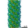 22701MC M: map data used to model this data C: citing same article ( |
|---|---|
| Similar structure data |


-Links
 PDBj
PDBj- Assembly
| Deposited unit | 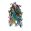
|
|---|---|
| 1 |
|
-Components
| #1: Protein | Mass: 20482.811 Da / Num. of mol.: 28 / Source method: isolated from a natural source Source: (natural) Strain: E2348/69 / EPEC / References: UniProt: B7UM94 |
|---|
-Experimental details
-Experiment
| Experiment | Method: ELECTRON MICROSCOPY |
|---|---|
| EM experiment | Aggregation state: FILAMENT / 3D reconstruction method: helical reconstruction |
- Sample preparation
| Component | Name: EspA filament / Type: ORGANELLE OR CELLULAR COMPONENT / Entity ID: all / Source: NATURAL |
|---|---|
| Source (natural) | Organism: |
| Buffer solution | pH: 7.4 |
| Specimen | Embedding applied: NO / Shadowing applied: NO / Staining applied: NO / Vitrification applied: YES |
| Vitrification | Instrument: FEI VITROBOT MARK IV / Cryogen name: ETHANE |
- Electron microscopy imaging
| Experimental equipment |  Model: Titan Krios / Image courtesy: FEI Company |
|---|---|
| Microscopy | Model: FEI TITAN KRIOS |
| Electron gun | Electron source:  FIELD EMISSION GUN / Accelerating voltage: 300 kV / Illumination mode: FLOOD BEAM FIELD EMISSION GUN / Accelerating voltage: 300 kV / Illumination mode: FLOOD BEAM |
| Electron lens | Mode: BRIGHT FIELD |
| Image recording | Electron dose: 0.475 e/Å2 / Film or detector model: FEI FALCON III (4k x 4k) |
- Processing
| EM software | Name: RELION / Version: 3 / Category: 3D reconstruction |
|---|---|
| CTF correction | Type: PHASE FLIPPING AND AMPLITUDE CORRECTION |
| Helical symmerty | Angular rotation/subunit: 64.3 ° / Axial rise/subunit: 4.4 Å / Axial symmetry: C1 |
| 3D reconstruction | Resolution: 3.56 Å / Resolution method: FSC 0.143 CUT-OFF / Num. of particles: 15523 / Symmetry type: HELICAL |
