 Movie
Movie Controller

[English] 日本語
 Yorodumi
- EMDB-22701: Structure of the EPEC type III secretion injectisome EspA filament -
+ Open data

- Basic information
| Entry | Database: EMDB / ID: EMD-22701 | |||||||||
|---|---|---|---|---|---|---|---|---|---|---|
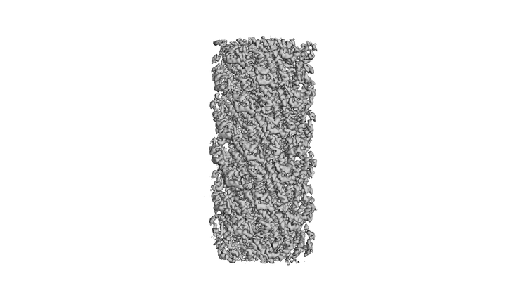
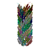
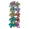
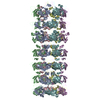
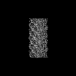
| Title | Structure of the EPEC type III secretion injectisome EspA filament | |||||||||
 Map data Map data | EPEC type III secretion injectisome EspA filament | |||||||||
 Sample Sample |
| |||||||||
 Keywords Keywords | Transport / filament / secretion system / PROTEIN TRANSPORT | |||||||||
| Function / homology | EspA-like secreted protein / EspA-like secreted protein / EspA/CesA-like / Translocon EspA Function and homology information Function and homology information | |||||||||
| Biological species |  | |||||||||
| Method | helical reconstruction / cryo EM / Resolution: 3.56 Å | |||||||||
 Authors Authors | Lyons BJE / Atkinson CE | |||||||||
| Funding support |  Canada, 2 items Canada, 2 items
| |||||||||
 Citation Citation | Journal: Structure / Year: 2021 Title: Cryo-EM structure of the EspA filament from enteropathogenic Escherichia coli: Revealing the mechanism of effector translocation in the T3SS. Authors: Bronwyn J E Lyons / Claire E Atkinson / Wanyin Deng / Antonio Serapio-Palacios / B Brett Finlay / Natalie C J Strynadka / Abstract: The type III secretion system (T3SS) is a virulence mechanism employed by Gram-negative pathogens. The T3SS forms a proteinaceous channel that projects a needle into the extracellular medium where it ...The type III secretion system (T3SS) is a virulence mechanism employed by Gram-negative pathogens. The T3SS forms a proteinaceous channel that projects a needle into the extracellular medium where it interacts with the host cell to deliver virulence factors. Enteropathogenic Escherichia coli (EPEC) is unique in adopting a needle extension to the T3SS-a filament formed by EspA-which is absolutely required for efficient colonization of the gut. Here, we describe the cryoelectron microscopy structure of native EspA filaments from EPEC at 3.6-Å resolution. Within the filament, positively charged residues adjacent to a hydrophobic groove line the lumen of the filament in a spiral manner, suggesting a mechanism of substrate translocation mediated via electrostatics. Using structure-guided mutagenesis, in vivo studies corroborate the role of these residues in secretion and translocation function. The high-resolution structure of the EspA filament could aid in structure-guided drug design of antivirulence therapeutics. | |||||||||
| History |
|
- Structure visualization
| Movie |
Movie viewer |
|---|---|
| Structure viewer | EM map: SurfViewMolmilJmol/JSmol |
| Supplemental images |
- Downloads & links
-EMDB archive
| Map data | emd_22701.map.gz | 5.8 MB | EMDB map data format | |
|---|---|---|---|---|
| Header (meta data) | emd-22701-v30.xmlemd-22701.xml | 9.5 KB 9.5 KB | Display Display | EMDB header |
| FSC (resolution estimation) | emd_22701_fsc.xml | 8.8 KB | Display | FSC data file |
| Images |  emd_22701.png emd_22701.png | 46.3 KB | ||
| Filedesc metadata | emd-22701.cif.gz | 4.7 KB | ||
| Archive directory |  http://ftp.pdbj.org/pub/emdb/structures/EMD-22701ftp://ftp.pdbj.org/pub/emdb/structures/EMD-22701 http://ftp.pdbj.org/pub/emdb/structures/EMD-22701ftp://ftp.pdbj.org/pub/emdb/structures/EMD-22701 | HTTPS FTP |
-Related structure data
| Related structure data | 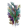 7k7kMC M: atomic model generated by this map C: citing same article ( |
|---|---|
| Similar structure data |

-Links
| EMDB pages | EMDB (EBI/PDBe) / EMDataResource |
|---|
-Map
| File | Download / File: emd_22701.map.gz / Format: CCP4 / Size: 59.6 MB / Type: IMAGE STORED AS FLOATING POINT NUMBER (4 BYTES) | ||||||||||||||||||||||||||||||||||||||||||||||||||||||||||||
|---|---|---|---|---|---|---|---|---|---|---|---|---|---|---|---|---|---|---|---|---|---|---|---|---|---|---|---|---|---|---|---|---|---|---|---|---|---|---|---|---|---|---|---|---|---|---|---|---|---|---|---|---|---|---|---|---|---|---|---|---|---|
| Annotation | EPEC type III secretion injectisome EspA filament | ||||||||||||||||||||||||||||||||||||||||||||||||||||||||||||
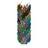
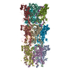
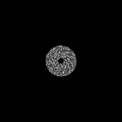
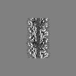
| Projections & slices | Image control
Images are generated by Spider. | ||||||||||||||||||||||||||||||||||||||||||||||||||||||||||||
| Voxel size | X=Y=Z: 1.75 Å | ||||||||||||||||||||||||||||||||||||||||||||||||||||||||||||
| Density |
| ||||||||||||||||||||||||||||||||||||||||||||||||||||||||||||
| Symmetry | Space group: 1 | ||||||||||||||||||||||||||||||||||||||||||||||||||||||||||||
| Details | EMDB XML:
CCP4 map header:
| ||||||||||||||||||||||||||||||||||||||||||||||||||||||||||||
 Z (Sec.)
 Y (Row.)
 X (Col.)


-Supplemental data
- Sample components
-Entire : EspA filament
| Entire | Name: EspA filament |
|---|---|
| Components |
|
-Supramolecule #1: EspA filament
| Supramolecule | Name: EspA filament / type: organelle_or_cellular_component / ID: 1 / Parent: 0 / Macromolecule list: all |
|---|---|
| Source (natural) | Organism: |
-Macromolecule #1: Translocon EspA
| Macromolecule | Name: Translocon EspA / type: protein_or_peptide / ID: 1 / Number of copies: 28 / Enantiomer: LEVO |
|---|---|
| Source (natural) | Organism: Strain: E2348/69 / EPEC |
| Molecular weight | Theoretical: 20.482811 KDa |
| Sequence | String: MDTSTTASVA SANASTSTSM AYDLGSMSKD DVIDLFNKLG VFQAAILMFA YMYQAQSDLS IAKFADMNEA SKESTTAQKM ANLVDAKIA DVQSSSDKNA KAQLPDEVIS YINDPRNDIT ISGIDNINAQ LGAGDLQTVK AAISAKANNL TTTVNNSQLE I QQMSNTLN ...String: MDTSTTASVA SANASTSTSM AYDLGSMSKD DVIDLFNKLG VFQAAILMFA YMYQAQSDLS IAKFADMNEA SKESTTAQKM ANLVDAKIA DVQSSSDKNA KAQLPDEVIS YINDPRNDIT ISGIDNINAQ LGAGDLQTVK AAISAKANNL TTTVNNSQLE I QQMSNTLN LLTSARSDMQ SLQYRTISGI SLGK UniProtKB: Translocon EspA |
-Experimental details
-Structure determination
| Method | cryo EM |
|---|---|
 Processing Processing | helical reconstruction |
| Aggregation state | filament |
-Sample preparation
| Buffer | pH: 7.4 |
|---|---|
| Vitrification | Cryogen name: ETHANE / Instrument: FEI VITROBOT MARK IV |
- Electron microscopy
| Microscope | FEI TITAN KRIOS |
|---|---|
| Image recording | Film or detector model: FEI FALCON III (4k x 4k) / Average electron dose: 0.475 e/Å2 |
| Electron beam | Acceleration voltage: 300 kV / Electron source:  FIELD EMISSION GUN FIELD EMISSION GUN |
| Electron optics | Illumination mode: FLOOD BEAM / Imaging mode: BRIGHT FIELD |
| Experimental equipment |  Model: Titan Krios / Image courtesy: FEI Company |
-Image processing
| Final reconstruction | Applied symmetry - Helical parameters - Δz: 4.4 Å Applied symmetry - Helical parameters - Δ&Phi: 64.3 ° Applied symmetry - Helical parameters - Axial symmetry: C1 (asymmetric) Resolution.type: BY AUTHOR / Resolution: 3.56 Å / Resolution method: FSC 0.143 CUT-OFF / Software - Name: RELION (ver. 3) / Number images used: 15523 |
|---|---|
| Startup model | Type of model: NONE |
| Final angle assignment | Type: NOT APPLICABLE |
| FSC plot (resolution estimation) |  |
