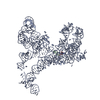
 Movie
Movie Controller


+ Open data

- Basic information
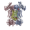
| Entry | Database: PDB / ID: 7d3e | |||||||||
|---|---|---|---|---|---|---|---|---|---|---|
| Title | Cryo-EM structure of human DUOX1-DUOXA1 in low-calcium state | |||||||||
 Components Components |
| |||||||||
 Keywords Keywords | ELECTRON TRANSPORT / DUOX / DUOXA / NOX / NADPH / FAD / Haem | |||||||||
| Function / homology |  Function and homology information Function and homology informationregulation of thyroid hormone generation / cuticle development / NAD(P)H oxidase (H2O2-forming) / Thyroxine biosynthesis / positive regulation of hydrogen peroxide biosynthetic process / hormone biosynthetic process / hydrogen peroxide metabolic process / NAD(P)H oxidase H2O2-forming activity / superoxide-generating NAD(P)H oxidase activity / NADPH oxidase complex ...regulation of thyroid hormone generation / cuticle development / NAD(P)H oxidase (H2O2-forming) / Thyroxine biosynthesis / positive regulation of hydrogen peroxide biosynthetic process / hormone biosynthetic process / hydrogen peroxide metabolic process / NAD(P)H oxidase H2O2-forming activity / superoxide-generating NAD(P)H oxidase activity / NADPH oxidase complex / thyroid hormone generation / Oxidoreductases / hydrogen peroxide biosynthetic process / superoxide anion generation / positive regulation of cell motility / positive regulation of wound healing / cell leading edge / Oxidoreductases; Acting on a peroxide as acceptor; Peroxidases / response to cAMP / NADPH binding / FAD binding / positive regulation of neuron differentiation / hydrogen peroxide catabolic process / peroxidase activity / defense response / cytokine-mediated signaling pathway / NADP binding / intracellular protein localization / protein transport / regulation of inflammatory response / response to oxidative stress / apical plasma membrane / protein heterodimerization activity / heme binding / calcium ion binding / endoplasmic reticulum membrane / enzyme binding / cell surface / endoplasmic reticulum / membrane / plasma membrane Similarity search - Function | |||||||||
| Biological species |  Homo sapiens (human) Homo sapiens (human) | |||||||||
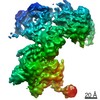
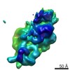
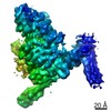
| Method | ELECTRON MICROSCOPY / single particle reconstruction / cryo EM / Resolution: 2.8 Å | |||||||||
 Authors Authors | Chen, L. / Wu, J.X. | |||||||||
| Funding support |  China, 2items China, 2items
| |||||||||
 Citation Citation | Journal: Nat Commun / Year: 2021 Title: Structures of human dual oxidase 1 complex in low-calcium and high-calcium states. Authors: Jing-Xiang Wu / Rui Liu / Kangcheng Song / Lei Chen / Abstract: Dual oxidases (DUOXs) produce hydrogen peroxide by transferring electrons from intracellular NADPH to extracellular oxygen. They are involved in many crucial biological processes and human diseases, ...Dual oxidases (DUOXs) produce hydrogen peroxide by transferring electrons from intracellular NADPH to extracellular oxygen. They are involved in many crucial biological processes and human diseases, especially in thyroid diseases. DUOXs are protein complexes co-assembled from the catalytic DUOX subunits and the auxiliary DUOXA subunits and their activities are regulated by intracellular calcium concentrations. Here, we report the cryo-EM structures of human DUOX1-DUOXA1 complex in both high-calcium and low-calcium states. These structures reveal the DUOX1 complex is a symmetric 2:2 hetero-tetramer stabilized by extensive inter-subunit interactions. Substrate NADPH and cofactor FAD are sandwiched between transmembrane domain and the cytosolic dehydrogenase domain of DUOX. In the presence of calcium ions, intracellular EF-hand modules might enhance the catalytic activity of DUOX by stabilizing the dehydrogenase domain in a conformation that allows electron transfer. | |||||||||
| History |
|
- Structure visualization
| Movie |
Movie viewer |
|---|---|
| Structure viewer | Molecule: MolmilJmol/JSmol |
- Downloads & links
-Download
| PDBx/mmCIF format | 7d3e.cif.gz | 679.3 KB | Display | PDBx/mmCIF format |
|---|---|---|---|---|
| PDB format | pdb7d3e.ent.gz | 532.3 KB | Display | PDB format |
| PDBx/mmJSON format | 7d3e.json.gz | Tree view | PDBx/mmJSON format | |
| Others |  Other downloads Other downloads |
-Validation report
| Arichive directory | https://data.pdbj.org/pub/pdb/validation_reports/d3/7d3eftp://data.pdbj.org/pub/pdb/validation_reports/d3/7d3e | HTTPS FTP |
|---|
-Related structure data
| Related structure data |  30555MC  7d3fC M: map data used to model this data C: citing same article ( |
|---|---|
| Similar structure data |


-Links
 PDBj
PDBj


- Assembly
| Deposited unit | 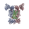
|
|---|---|
| 1 |
|
-Components
-Protein , 2 types, 4 molecules ACBD
| #1: Protein | Mass: 177483.828 Da / Num. of mol.: 2 Source method: isolated from a genetically manipulated source Source: (gene. exp.) Homo sapiens (human) / Gene: DUOX1, DUOX, LNOX1, THOX1 / Production host: Homo sapiens (human)References: UniProt: Q9NRD9, Oxidoreductases; Acting on a peroxide as acceptor; Peroxidases, NAD(P)H oxidase (H2O2-forming) #2: Protein | Mass: 53579.188 Da / Num. of mol.: 2 Source method: isolated from a genetically manipulated source Source: (gene. exp.) Homo sapiens (human) / Gene: DUOXA1, NIP, NUMBIP / Production host: Homo sapiens (human) / References: UniProt: Q1HG43 |
|---|
-Sugars , 2 types, 12 molecules 
| #3: Polysaccharide | | #7: Sugar | ChemComp-NAG /  Type: D-saccharide, beta linking / Mass: 221.208 Da / Num. of mol.: 10 / Source method: isolated from a natural source / Formula: C8H15NO6 Type: D-saccharide, beta linking / Mass: 221.208 Da / Num. of mol.: 10 / Source method: isolated from a natural source / Formula: C8H15NO6 |
|---|
-Non-polymers , 5 types, 14 molecules 


| #4: Chemical | ChemComp-HEM /  Mass: 616.487 Da / Num. of mol.: 4 / Source method: obtained synthetically / Formula: C34H32FeN4O4 / Feature type: SUBJECT OF INVESTIGATION Mass: 616.487 Da / Num. of mol.: 4 / Source method: obtained synthetically / Formula: C34H32FeN4O4 / Feature type: SUBJECT OF INVESTIGATION#5: Chemical |  Mass: 745.421 Da / Num. of mol.: 2 / Source method: obtained synthetically / Formula: C21H30N7O17P3 / Feature type: SUBJECT OF INVESTIGATION Mass: 745.421 Da / Num. of mol.: 2 / Source method: obtained synthetically / Formula: C21H30N7O17P3 / Feature type: SUBJECT OF INVESTIGATION#6: Chemical |  Mass: 785.550 Da / Num. of mol.: 2 / Source method: isolated from a natural source / Formula: C27H33N9O15P2 / Feature type: SUBJECT OF INVESTIGATION / Comment: FAD*YM Mass: 785.550 Da / Num. of mol.: 2 / Source method: isolated from a natural source / Formula: C27H33N9O15P2 / Feature type: SUBJECT OF INVESTIGATION / Comment: FAD*YM#8: Chemical | ChemComp-NA /  Mass: 22.990 Da / Num. of mol.: 4 / Source method: isolated from a natural source / Formula: Na Mass: 22.990 Da / Num. of mol.: 4 / Source method: isolated from a natural source / Formula: Na#9: Water | ChemComp-HOH / | Mass: 18.015 Da / Num. of mol.: 2 / Source method: isolated from a natural source / Formula: H2O |
|---|
-Details
| Has ligand of interest | Y |
|---|---|
| Has protein modification | Y |
-Experimental details
-Experiment
| Experiment | Method: ELECTRON MICROSCOPY |
|---|---|
| EM experiment | Aggregation state: PARTICLE / 3D reconstruction method: single particle reconstruction |
- Sample preparation
| Component | Name: human DUOX1-DUOXA1 complex / Type: COMPLEX / Entity ID: #1-#2 / Source: RECOMBINANT |
|---|---|
| Source (natural) | Organism: Homo sapiens (human) |
| Source (recombinant) | Organism: Homo sapiens (human) |
| Buffer solution | pH: 7 |
| Specimen | Embedding applied: NO / Shadowing applied: NO / Staining applied: NO / Vitrification applied: YES |
| Vitrification | Cryogen name: ETHANE |
- Electron microscopy imaging
| Experimental equipment |  Model: Titan Krios / Image courtesy: FEI Company |
|---|---|
| Microscopy | Model: FEI TITAN KRIOS |
| Electron gun | Electron source:  FIELD EMISSION GUN / Accelerating voltage: 300 kV / Illumination mode: FLOOD BEAM FIELD EMISSION GUN / Accelerating voltage: 300 kV / Illumination mode: FLOOD BEAM |
| Electron lens | Mode: BRIGHT FIELD |
| Image recording | Electron dose: 48 e/Å2 / Film or detector model: GATAN K2 SUMMIT (4k x 4k) |
- Processing
| CTF correction | Type: NONE |
|---|---|
| 3D reconstruction | Resolution: 2.8 Å / Resolution method: FSC 0.143 CUT-OFF / Num. of particles: 125984 / Symmetry type: POINT |
