 Movie
Movie Controller


+ Open data

- Basic information
| Entry | Database: PDB / ID: 7cbp | ||||||||||||||||||||||||||||||
|---|---|---|---|---|---|---|---|---|---|---|---|---|---|---|---|---|---|---|---|---|---|---|---|---|---|---|---|---|---|---|---|
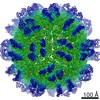
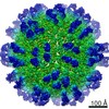
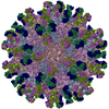
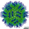
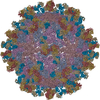
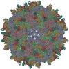
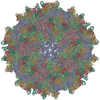
| Title | CryoEM structure of Zika virus with Fab at 4.1 Angstrom | ||||||||||||||||||||||||||||||
 Components Components |
| ||||||||||||||||||||||||||||||
 Keywords Keywords | VIRUS / Zika virus / Fab antibody | ||||||||||||||||||||||||||||||
| Function / homology |  Function and homology information Function and homology informationflavivirin / symbiont-mediated suppression of host JAK-STAT cascade via inhibition of host TYK2 activity / symbiont-mediated suppression of host JAK-STAT cascade via inhibition of STAT2 activity / symbiont-mediated suppression of host JAK-STAT cascade via inhibition of STAT1 activity / negative regulation of innate immune response / viral capsid / nucleoside-triphosphate phosphatase / double-stranded RNA binding / 4 iron, 4 sulfur cluster binding / clathrin-dependent endocytosis of virus by host cell ...flavivirin / symbiont-mediated suppression of host JAK-STAT cascade via inhibition of host TYK2 activity / symbiont-mediated suppression of host JAK-STAT cascade via inhibition of STAT2 activity / symbiont-mediated suppression of host JAK-STAT cascade via inhibition of STAT1 activity / negative regulation of innate immune response / viral capsid / nucleoside-triphosphate phosphatase / double-stranded RNA binding / 4 iron, 4 sulfur cluster binding / clathrin-dependent endocytosis of virus by host cell / mRNA (guanine-N7)-methyltransferase / molecular adaptor activity / methyltransferase cap1 / methyltransferase cap1 activity / mRNA 5'-cap (guanine-N7-)-methyltransferase activity / RNA helicase activity / protein dimerization activity / host cell perinuclear region of cytoplasm / host cell endoplasmic reticulum membrane / RNA helicase / symbiont-mediated suppression of host type I interferon-mediated signaling pathway / serine-type endopeptidase activity / symbiont-mediated activation of host autophagy / RNA-directed RNA polymerase / viral RNA genome replication / RNA-directed RNA polymerase activity / fusion of virus membrane with host endosome membrane / viral envelope / centrosome / lipid binding / virion attachment to host cell / GTP binding / host cell nucleus / virion membrane / structural molecule activity / ATP hydrolysis activity / proteolysis / extracellular region / ATP binding / metal ion binding Similarity search - Function | ||||||||||||||||||||||||||||||
| Biological species |  Homo sapiens (human) Homo sapiens (human)  Zika virus Zika virus | ||||||||||||||||||||||||||||||
| Method | ELECTRON MICROSCOPY / single particle reconstruction / cryo EM / Resolution: 4.1 Å | ||||||||||||||||||||||||||||||
 Authors Authors | Tyagi, A. / Ahmed, T. / Shi, J. / Bhushan, S. | ||||||||||||||||||||||||||||||
| Funding support | 1items
| ||||||||||||||||||||||||||||||
 Citation Citation | Journal: J Struct Biol X / Year: 2020 Title: A complex between the Zika virion and the Fab of a broadly cross-reactive neutralizing monoclonal antibody revealed by cryo-EM and single particle analysis at 4.1 Å resolution. Authors: Anu Tyagi / Tofayel Ahmed / Jian Shi / Shashi Bhushan /  Abstract: Zika virus (ZIKV) recently emerged as a major public health concern because it can cause fetal microcephaly and neurological disease such as the Guillain-Barré syndrome. A particularly potent class ...Zika virus (ZIKV) recently emerged as a major public health concern because it can cause fetal microcephaly and neurological disease such as the Guillain-Barré syndrome. A particularly potent class of broadly neutralizing antibodies (nAbs) targets a quaternary epitope located at the interface of two envelope proteins monomers, exposed at the surface of the mature virion. This "E-dimer-dependent epitope" (EDE), comprises the fusion loop of one monomer at the tip of domain II of E and a portion of the domains I and III of the adjacent monomer. Since this epitope largely overlaps with the binding site of the precursor membrane protein (prM) during Zika virion maturation, its molecular surface is evolutionary conserved in flaviviruses such as Dengue and Zika viruses, and can elicit antibodies that broadly neutralize various ZIKV strains. Here, we present a cryo-EM reconstruction at 4.1 Å resolution of the virion bound to the antigen binding fragment (Fab) of an antibody that targets this mutationally-constrained quaternary epitope. The Fab incompletely covers the surface of the virion as it does not bind next to its 5-fold icosahedral axes. The structure reveals details of the binding mode of this potent neutralizing class of antibodies and can inform the design of immunogens and vaccines targeting this conserved epitope. | ||||||||||||||||||||||||||||||
| History |
|
- Structure visualization
| Movie |
Movie viewer |
|---|---|
| Structure viewer | Molecule: MolmilJmol/JSmol |
- Downloads & links
-Download
| PDBx/mmCIF format | 7cbp.cif.gz | 439.2 KB | Display | PDBx/mmCIF format |
|---|---|---|---|---|
| PDB format | pdb7cbp.ent.gz | 363 KB | Display | PDB format |
| PDBx/mmJSON format | 7cbp.json.gz | Tree view | PDBx/mmJSON format | |
| Others |  Other downloads Other downloads |
-Validation report
| Arichive directory | https://data.pdbj.org/pub/pdb/validation_reports/cb/7cbpftp://data.pdbj.org/pub/pdb/validation_reports/cb/7cbp | HTTPS FTP |
|---|
-Related structure data
| Related structure data |  30337MC M: map data used to model this data C: citing same article ( |
|---|---|
| Similar structure data |

-Links
 PDBj
PDBj


- Assembly
| Deposited unit | 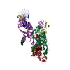
|
|---|---|
| 1 | x 60
|
| 2 |
|
| 3 | x 5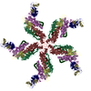
|
| 4 | x 6
|
| 5 | 
|
| Symmetry | Point symmetry: (Schoenflies symbol: I (icosahedral)) |
-Components
| #1: Protein | Mass: 8496.883 Da / Num. of mol.: 3 / Source method: isolated from a natural source / Source: (natural) Zika virus / References: UniProt: A0A024B7W1#2: Protein | Mass: 54186.762 Da / Num. of mol.: 3 / Source method: isolated from a natural source / Source: (natural) Zika virus / References: UniProt: A0A024B7W1#3: Antibody | Mass: 23612.326 Da / Num. of mol.: 2 Source method: isolated from a genetically manipulated source Source: (gene. exp.) Homo sapiens (human) / Production host: Homo sapiens (human)#4: Antibody | Mass: 23818.539 Da / Num. of mol.: 2 Source method: isolated from a genetically manipulated source Source: (gene. exp.) Homo sapiens (human) / Production host: Homo sapiens (human)#5: Polysaccharide | 2-acetamido-2-deoxy-beta-D-glucopyranose-(1-4)-2-acetamido-2-deoxy-beta-D-glucopyranose | Source method: isolated from a genetically manipulated source Has ligand of interest | N | Has protein modification | Y | |
|---|
-Experimental details
-Experiment
| Experiment | Method: ELECTRON MICROSCOPY |
|---|---|
| EM experiment | Aggregation state: PARTICLE / 3D reconstruction method: single particle reconstruction |
- Sample preparation
| Component |
| ||||||||||||||||||||||||
|---|---|---|---|---|---|---|---|---|---|---|---|---|---|---|---|---|---|---|---|---|---|---|---|---|---|
| Source (natural) |
| ||||||||||||||||||||||||
| Details of virus | Empty: YES / Enveloped: YES / Isolate: OTHER / Type: VIRION | ||||||||||||||||||||||||
| Buffer solution | pH: 8 | ||||||||||||||||||||||||
| Specimen | Embedding applied: NO / Shadowing applied: NO / Staining applied: NO / Vitrification applied: YES | ||||||||||||||||||||||||
| Vitrification | Cryogen name: ETHANE |
- Electron microscopy imaging
| Experimental equipment |  Model: Titan Krios / Image courtesy: FEI Company |
|---|---|
| Microscopy | Model: FEI TITAN KRIOS |
| Electron gun | Electron source:  FIELD EMISSION GUN / Accelerating voltage: 300 kV / Illumination mode: FLOOD BEAM FIELD EMISSION GUN / Accelerating voltage: 300 kV / Illumination mode: FLOOD BEAM |
| Electron lens | Mode: BRIGHT FIELD |
| Image recording | Electron dose: 48 e/Å2 / Film or detector model: FEI FALCON II (4k x 4k) |
- Processing
| EM software | Name: RELION / Category: classification |
|---|---|
| CTF correction | Type: PHASE FLIPPING ONLY |
| 3D reconstruction | Resolution: 4.1 Å / Resolution method: FSC 0.143 CUT-OFF / Num. of particles: 4610 / Symmetry type: POINT |
