 Movie
Movie Controller


+ Open data

- Basic information
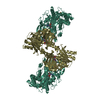
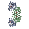
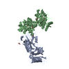
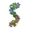
| Entry | Database: PDB / ID: 7ad3 | |||||||||
|---|---|---|---|---|---|---|---|---|---|---|
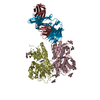
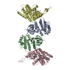
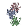
| Title | Class D GPCR Ste2 dimer coupled to two G proteins | |||||||||
 Components Components |
| |||||||||
 Keywords Keywords | MEMBRANE PROTEIN / Fungal GPCR / Dimer / Complex / Class D / Active State | |||||||||
| Function / homology |  Function and homology information Function and homology informationprotein localization to mating projection tip / PLC beta mediated events / : / Acetylcholine regulates insulin secretion / G alpha (q) signalling events / ADP signalling through P2Y purinoceptor 1 / : / mating projection / G-protein beta/gamma-subunit complex / : ...protein localization to mating projection tip / PLC beta mediated events / : / Acetylcholine regulates insulin secretion / G alpha (q) signalling events / ADP signalling through P2Y purinoceptor 1 / : / mating projection / G-protein beta/gamma-subunit complex / : / chemotropism / Cdc24p-Far1p-Gbetagamma complex / CDC42 GTPase cycle / G alpha (12/13) signalling events / mating pheromone activity / mating-type factor pheromone receptor activity / nuclear migration involved in conjugation with cellular fusion / High laminar flow shear stress activates signaling by PIEZO1 and PECAM1:CDH5:KDR in endothelial cells / response to pheromone triggering conjugation with cellular fusion / G protein-coupled receptor homodimeric complex / karyogamy involved in conjugation with cellular fusion / regulation of carbohydrate metabolic process / Cooperation of PDCL (PhLP1) and TRiC/CCT in G-protein beta folding / establishment of protein localization to plasma membrane / mating / : / G-protein gamma-subunit binding / invasive growth in response to glucose limitation / cupric ion binding / G-protein alpha-subunit binding / cell periphery / G protein-coupled receptor binding / small GTPase binding / G-protein beta/gamma-subunit complex binding / heterotrimeric G-protein complex / G-protein beta-subunit binding / signaling receptor complex adaptor activity / scaffold protein binding / cell cortex / endosome membrane / endosome / G protein-coupled receptor signaling pathway / GTPase activity / protein kinase binding / GTP binding / signal transduction / extracellular region / metal ion binding / plasma membrane / cytoplasm / cytosol Similarity search - Function | |||||||||
| Biological species |  | |||||||||
| Method | ELECTRON MICROSCOPY / single particle reconstruction / cryo EM / Resolution: 3.5 Å | |||||||||
 Authors Authors | Velazhahan, V. / Tate, C. | |||||||||
| Funding support |  United Kingdom, 2items United Kingdom, 2items
| |||||||||
 Citation Citation | Journal: Nature / Year: 2021 Title: Structure of the class D GPCR Ste2 dimer coupled to two G proteins. Authors: Vaithish Velazhahan / Ning Ma / Gáspár Pándy-Szekeres / Albert J Kooistra / Yang Lee / David E Gloriam / Nagarajan Vaidehi / Christopher G Tate /    Abstract: G-protein-coupled receptors (GPCRs) are divided phylogenetically into six classes, denoted A to F. More than 370 structures of vertebrate GPCRs (belonging to classes A, B, C and F) have been ...G-protein-coupled receptors (GPCRs) are divided phylogenetically into six classes, denoted A to F. More than 370 structures of vertebrate GPCRs (belonging to classes A, B, C and F) have been determined, leading to a substantial understanding of their function. By contrast, there are no structures of class D GPCRs, which are found exclusively in fungi where they regulate survival and reproduction. Here we determine the structure of a class D GPCR, the Saccharomyces cerevisiae pheromone receptor Ste2, in an active state coupled to the heterotrimeric G protein Gpa1-Ste4-Ste18. Ste2 was purified as a homodimer coupled to two G proteins. The dimer interface of Ste2 is formed by the N terminus, the transmembrane helices H1, H2 and H7, and the first extracellular loop ECL1. We establish a class D1 generic residue numbering system (CD1) to enable comparisons with orthologues and with other GPCR classes. The structure of Ste2 bears similarities in overall topology to class A GPCRs, but the transmembrane helix H4 is shifted by more than 20 Å and the G-protein-binding site is a shallow groove rather than a cleft. The structure provides a template for the design of novel drugs to target fungal GPCRs, which could be used to treat numerous intractable fungal diseases. | |||||||||
| History |
|
- Structure visualization
| Movie |
Movie viewer |
|---|---|
| Structure viewer | Molecule: MolmilJmol/JSmol |
 UCSF Chimera
- Downloads & links
-Download
| PDBx/mmCIF format | 7ad3.cif.gz | 234 KB | Display | PDBx/mmCIF format |
|---|---|---|---|---|
| PDB format | pdb7ad3.ent.gz | 178.3 KB | Display | PDB format |
| PDBx/mmJSON format | 7ad3.json.gz | Tree view | PDBx/mmJSON format | |
| Others |  Other downloads Other downloads |
-Validation report
| Arichive directory | https://data.pdbj.org/pub/pdb/validation_reports/ad/7ad3ftp://data.pdbj.org/pub/pdb/validation_reports/ad/7ad3 | HTTPS FTP |
|---|
-Related structure data
| Related structure data |  11720MC  7qa8C  7qb9C  7qbcC  7qbiC M: map data used to model this data C: citing same article ( |
|---|---|
| Similar structure data | |
| EM raw data | EMPIAR-10550 (Title: Structure of the class D GPCR Ste2 dimer coupled to two G proteins Data size: 6.4 TB / Data #1: LMB Krios1 Movies [micrographs - multiframe] / Data #2: LMB Krios2 Movies [micrographs - multiframe] / Data #3: eBic Krios1 Movies [micrographs - multiframe]) |


-Links
 PDBj
PDBj


- Assembly
| Deposited unit | 
|
|---|---|
| 1 |
|
-Components
-Protein , 2 types, 3 molecules BAF
| #1: Protein | Mass: 47885.402 Da / Num. of mol.: 2 Source method: isolated from a genetically manipulated source Source: (gene. exp.) Gene: STE2 / Production host:  Trichoplusia ni (cabbage looper) / References: UniProt: P0CI39 Trichoplusia ni (cabbage looper) / References: UniProt: P0CI39#3: Protein | | Mass: 46626.953 Da / Num. of mol.: 1 Source method: isolated from a genetically manipulated source Source: (gene. exp.) Gene: STE4, GI526_G0005548 / Production host: Trichoplusia ni (cabbage looper) / References: UniProt: A0A6A5Q727, UniProt: P18851*PLUS |
|---|
-Guanine nucleotide-binding protein ... , 2 types, 3 molecules EHG
| #4: Protein | Mass: 26316.305 Da / Num. of mol.: 2 Source method: isolated from a genetically manipulated source Source: (gene. exp.) Strain: ATCC 204508 / S288c / Gene: GPA1, CDC70, DAC1, SCG1, YHR005C Production host:  References: UniProt: P08539 #5: Protein | | Mass: 12477.051 Da / Num. of mol.: 1 Source method: isolated from a genetically manipulated source Source: (gene. exp.) Gene: STE18, GI526_G0003318, PACBIOSEQ_LOCUS2168, PACBIOSEQ_LOCUS3627, PACBIOSEQ_LOCUS3692, PACBIOSEQ_LOCUS3742, PACBIOSEQ_LOCUS3767 Production host: Trichoplusia ni (cabbage looper) / References: UniProt: A0A6A5PT44, UniProt: P18852*PLUS |
|---|
-Protein/peptide / Sugars / Non-polymers , 3 types, 10 molecules KI

| #2: Protein/peptide | Mass: 1685.986 Da / Num. of mol.: 2 / Source method: obtained synthetically / Source: (synth.) #6: Sugar |  Type: D-saccharide, beta linking / Mass: 221.208 Da / Num. of mol.: 2 / Source method: obtained synthetically / Formula: C8H15NO6 Type: D-saccharide, beta linking / Mass: 221.208 Da / Num. of mol.: 2 / Source method: obtained synthetically / Formula: C8H15NO6#7: Chemical | ChemComp-Y01 /  Mass: 486.726 Da / Num. of mol.: 6 / Source method: obtained synthetically / Formula: C31H50O4 Mass: 486.726 Da / Num. of mol.: 6 / Source method: obtained synthetically / Formula: C31H50O4 |
|---|
-Details
| Has ligand of interest | N |
|---|---|
| Has protein modification | Y |
-Experimental details
-Experiment
| Experiment | Method: ELECTRON MICROSCOPY |
|---|---|
| EM experiment | Aggregation state: PARTICLE / 3D reconstruction method: single particle reconstruction |
- Sample preparation
| Component |
| ||||||||||||||||||||||||||||||
|---|---|---|---|---|---|---|---|---|---|---|---|---|---|---|---|---|---|---|---|---|---|---|---|---|---|---|---|---|---|---|---|
| Molecular weight | Value: 0.2667 MDa / Experimental value: YES | ||||||||||||||||||||||||||||||
| Source (natural) |
| ||||||||||||||||||||||||||||||
| Source (recombinant) |
| ||||||||||||||||||||||||||||||
| Buffer solution | pH: 7.5 / Details: Solutions were made fresh and filtered | ||||||||||||||||||||||||||||||
| Buffer component |
| ||||||||||||||||||||||||||||||
| Specimen | Conc.: 0.7 mg/ml / Embedding applied: NO / Shadowing applied: NO / Staining applied: NO / Vitrification applied: YES / Details: The sample was purified as a monodisperse complex | ||||||||||||||||||||||||||||||
| Specimen support | Grid material: GOLD / Grid mesh size: 300 divisions/in. / Grid type: Quantifoil R1.2/1.3 | ||||||||||||||||||||||||||||||
| Vitrification | Instrument: FEI VITROBOT MARK IV / Cryogen name: ETHANE / Humidity: 100 % / Chamber temperature: 277 K |
- Electron microscopy imaging
| Experimental equipment |  Model: Titan Krios / Image courtesy: FEI Company | ||||||||||||||||||||
|---|---|---|---|---|---|---|---|---|---|---|---|---|---|---|---|---|---|---|---|---|---|
| EM imaging | Accelerating voltage: 300 kV / Calibrated defocus max: 2700 nm / Calibrated defocus min: 900 nm / Cryogen: NITROGEN / Electron source:
| ||||||||||||||||||||
| Image recording |
| ||||||||||||||||||||
| EM imaging optics |
|
- Processing
| Software | Name: PHENIX / Version: 1.17.1_3660: / Classification: refinement | ||||||||||||||||||||||||||||||||||||||||||||||||||||||||||||||||||||||
|---|---|---|---|---|---|---|---|---|---|---|---|---|---|---|---|---|---|---|---|---|---|---|---|---|---|---|---|---|---|---|---|---|---|---|---|---|---|---|---|---|---|---|---|---|---|---|---|---|---|---|---|---|---|---|---|---|---|---|---|---|---|---|---|---|---|---|---|---|---|---|---|
| EM software |
| ||||||||||||||||||||||||||||||||||||||||||||||||||||||||||||||||||||||
| Image processing |
| ||||||||||||||||||||||||||||||||||||||||||||||||||||||||||||||||||||||
| CTF correction |
| ||||||||||||||||||||||||||||||||||||||||||||||||||||||||||||||||||||||
| Particle selection |
| ||||||||||||||||||||||||||||||||||||||||||||||||||||||||||||||||||||||
| Symmetry |
| ||||||||||||||||||||||||||||||||||||||||||||||||||||||||||||||||||||||
| 3D reconstruction | Entry-ID: 7AD3 / Resolution method: FSC 0.143 CUT-OFF / Symmetry type: POINT
| ||||||||||||||||||||||||||||||||||||||||||||||||||||||||||||||||||||||
| Atomic model building | B value: 112 / Space: REAL / Target criteria: Correlation coefficient Details: Manual building was performed in Coot iterated with real space refinement in PHENIX. | ||||||||||||||||||||||||||||||||||||||||||||||||||||||||||||||||||||||
| Atomic model building | 3D fitting-ID: 1 / Accession code: 6G79 / Initial refinement model-ID: 1 / PDB-ID: 6G79 / Source name: PDB / Type: experimental model
| ||||||||||||||||||||||||||||||||||||||||||||||||||||||||||||||||||||||
| Refine LS restraints |
|

