 Movie
Movie Controller

[English] 日本語
 Yorodumi
- PDB-7z9p: The novel DNA binding mechanism of ridinilazole, a precision Clos... -
+ Open data

- Basic information
| Entry | Database: PDB / ID: 7z9p | ||||||||||||||||||||||||||||
|---|---|---|---|---|---|---|---|---|---|---|---|---|---|---|---|---|---|---|---|---|---|---|---|---|---|---|---|---|---|
| Title | The novel DNA binding mechanism of ridinilazole, a precision Clostridiodes difficile antibiotic | ||||||||||||||||||||||||||||
 Components Components | DNA (5'-D(P* Keywords KeywordsDNA / Clostridiodes difficile / antibiotic / DNA-binding / drug discovery / target identification | Function / homology | Ridinilazole / DNA / DNA (> 10) |  Function and homology information Function and homology informationBiological species | synthetic construct (others) | Method |  X-RAY DIFFRACTION / SYNCHROTRON / MOLECULAR REPLACEMENT / Resolution: 2.2 Å X-RAY DIFFRACTION / SYNCHROTRON / MOLECULAR REPLACEMENT / Resolution: 2.2 Å  Authors AuthorsMason, S. / Leonard, P.M. | Funding support | 1items |
 CitationJournal: Antimicrob.Agents Chemother. / Year: 2023 CitationJournal: Antimicrob.Agents Chemother. / Year: 2023Title: The Novel DNA Binding Mechanism of Ridinilazole, a Precision Clostridiodes difficile Antibiotic. Authors: Mason, C.S. / Avis, T. / Hu, C. / Nagalingam, N. / Mudaliar, M. / Coward, C. / Begum, K. / Gajewski, K. / Alam, M.J. / Basseres, E. / Moss, S. / Reich, S. / Duperchy, E. / Fox, K.R. / Garey, K.W. / Powell, D.J. History |
|
- Structure visualization
| Structure viewer | Molecule: MolmilJmol/JSmol |
|---|
- Downloads & links
-Download
| PDBx/mmCIF format | 7z9p.cif.gz | 53 KB | Display | PDBx/mmCIF format |
|---|---|---|---|---|
| PDB format | pdb7z9p.ent.gz | 36.6 KB | Display | PDB format |
| PDBx/mmJSON format | 7z9p.json.gz | Tree view | PDBx/mmJSON format | |
| Others |  Other downloads Other downloads |
-Validation report
| Arichive directory | https://data.pdbj.org/pub/pdb/validation_reports/z9/7z9pftp://data.pdbj.org/pub/pdb/validation_reports/z9/7z9p | HTTPS FTP |
|---|
-Related structure data
| Related structure data |  3u2nS S: Starting model for refinement |
|---|---|
| Similar structure data |
-Links
 PDBj
PDBj


- Assembly
| Deposited unit | 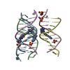
| ||||||||
|---|---|---|---|---|---|---|---|---|---|
| 1 | 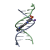
| ||||||||
| 2 | 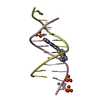
| ||||||||
| 3 | 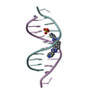
| ||||||||
| Unit cell |
|
-Components
-DNA chain , 1 types, 6 molecules ABCDEF
| #1: DNA chain | Mass: 3663.392 Da / Num. of mol.: 6 / Source method: obtained synthetically / Source: (synth.) synthetic construct (others) |
|---|
-Non-polymers , 5 types, 60 molecules 


| #2: Chemical |  Mass: 388.424 Da / Num. of mol.: 3 / Source method: obtained synthetically / Formula: C24H16N6 / Feature type: SUBJECT OF INVESTIGATION Mass: 388.424 Da / Num. of mol.: 3 / Source method: obtained synthetically / Formula: C24H16N6 / Feature type: SUBJECT OF INVESTIGATION#3: Chemical | ChemComp-SO4 /  Mass: 96.063 Da / Num. of mol.: 5 / Source method: obtained synthetically / Formula: SO4 Mass: 96.063 Da / Num. of mol.: 5 / Source method: obtained synthetically / Formula: SO4#4: Chemical | ChemComp-NA / |  Mass: 22.990 Da / Num. of mol.: 1 / Source method: obtained synthetically / Formula: Na Mass: 22.990 Da / Num. of mol.: 1 / Source method: obtained synthetically / Formula: Na#5: Chemical |  Mass: 65.409 Da / Num. of mol.: 3 / Source method: obtained synthetically / Formula: Zn Mass: 65.409 Da / Num. of mol.: 3 / Source method: obtained synthetically / Formula: Zn#6: Water | ChemComp-HOH / | Mass: 18.015 Da / Num. of mol.: 48 / Source method: isolated from a natural source / Formula: H2O |
|---|
-Details
| Has ligand of interest | Y |
|---|
-Experimental details
-Experiment
| Experiment | Method: X-RAY DIFFRACTION / Number of used crystals: 1 |
|---|
- Sample preparation
| Crystal | Density Matthews: 2.14 Å3/Da / Density % sol: 42.52 % |
|---|---|
| Crystal grow | Temperature: 287 K / Method: vapor diffusion, sitting drop / pH: 6.5 Details: 0.01 M Zinc sulphate heptahydrate 0.1 M MES 6.5 25 % v/v PEG 500 MME |
-Data collection
| Diffraction | Mean temperature: 100 K / Serial crystal experiment: N |
|---|---|
| Diffraction source | Source: SYNCHROTRON / Site: Diamond  / Beamline: I04 / Wavelength: 0.9795 Å / Beamline: I04 / Wavelength: 0.9795 Å |
| Detector | Type: DECTRIS EIGER2 XE 16M / Detector: PIXEL / Date: Nov 26, 2020 |
| Radiation | Protocol: SINGLE WAVELENGTH / Monochromatic (M) / Laue (L): M / Scattering type: x-ray |
| Radiation wavelength | Wavelength: 0.9795 Å / Relative weight: 1 |
| Reflection | Resolution: 2.2→30.08 Å / Num. obs: 8926 / % possible obs: 96.4 % / Redundancy: 1.7 % / CC1/2: 0.99 / Net I/σ(I): 5.3 |
| Reflection shell | Resolution: 2.2→2.27 Å / Mean I/σ(I) obs: 1.1 / Num. unique obs: 725 / CC1/2: 0.548 |
- Processing
| Software |
| ||||||||||||||||||||||||||||||||||||||||||||||||||||||||||||||||||||||||||||||||||||||||||||||||||||||||||||||||||||||||||||||||||||||||||||||||||||||||||||||||||||||||||||||||||||||
|---|---|---|---|---|---|---|---|---|---|---|---|---|---|---|---|---|---|---|---|---|---|---|---|---|---|---|---|---|---|---|---|---|---|---|---|---|---|---|---|---|---|---|---|---|---|---|---|---|---|---|---|---|---|---|---|---|---|---|---|---|---|---|---|---|---|---|---|---|---|---|---|---|---|---|---|---|---|---|---|---|---|---|---|---|---|---|---|---|---|---|---|---|---|---|---|---|---|---|---|---|---|---|---|---|---|---|---|---|---|---|---|---|---|---|---|---|---|---|---|---|---|---|---|---|---|---|---|---|---|---|---|---|---|---|---|---|---|---|---|---|---|---|---|---|---|---|---|---|---|---|---|---|---|---|---|---|---|---|---|---|---|---|---|---|---|---|---|---|---|---|---|---|---|---|---|---|---|---|---|---|---|---|---|
| Refinement | Method to determine structure: MOLECULAR REPLACEMENT Starting model: 3U2N Resolution: 2.2→30.1 Å / Cor.coef. Fo:Fc: 0.944 / Cor.coef. Fo:Fc free: 0.923 / SU B: 8.006 / SU ML: 0.207 / Cross valid method: THROUGHOUT / ESU R: 0.404 / ESU R Free: 0.231 / Stereochemistry target values: MAXIMUM LIKELIHOOD / Details: HYDROGENS HAVE BEEN ADDED IN THE RIDING POSITIONS
| ||||||||||||||||||||||||||||||||||||||||||||||||||||||||||||||||||||||||||||||||||||||||||||||||||||||||||||||||||||||||||||||||||||||||||||||||||||||||||||||||||||||||||||||||||||||
| Solvent computation | Ion probe radii: 0.8 Å / Shrinkage radii: 0.8 Å / VDW probe radii: 1.2 Å / Solvent model: MASK | ||||||||||||||||||||||||||||||||||||||||||||||||||||||||||||||||||||||||||||||||||||||||||||||||||||||||||||||||||||||||||||||||||||||||||||||||||||||||||||||||||||||||||||||||||||||
| Displacement parameters | Biso mean: 38.28 Å2
| ||||||||||||||||||||||||||||||||||||||||||||||||||||||||||||||||||||||||||||||||||||||||||||||||||||||||||||||||||||||||||||||||||||||||||||||||||||||||||||||||||||||||||||||||||||||
| Refinement step | Cycle: 1 / Resolution: 2.2→30.1 Å
| ||||||||||||||||||||||||||||||||||||||||||||||||||||||||||||||||||||||||||||||||||||||||||||||||||||||||||||||||||||||||||||||||||||||||||||||||||||||||||||||||||||||||||||||||||||||
| Refine LS restraints |
|