 Movie
Movie Controller


+ Open data

- Basic information
| Entry | Database: PDB / ID: 7yi7 | ||||||
|---|---|---|---|---|---|---|---|
| Title | Crystal structure of Human HPSE1 in complex with inhibitor | ||||||
 Components Components |
| ||||||
 Keywords Keywords | HYDROLASE/INHIBITOR / Endo-glucoronidase / Heparanase-1 / HEP / HPA / HPA1 / HPR1 / HPSE1 / HSE1 / HYDROLASE / HYDROLASE-INHIBITOR complex | ||||||
| Function / homology |  Function and homology information Function and homology informationheparanase / heparanase activity / regulation of hair follicle development / heparin metabolic process / proteoglycan metabolic process / heparan sulfate proteoglycan catabolic process / beta-glucuronidase activity / positive regulation of hair follicle development / HS-GAG degradation / protein transmembrane transport ...heparanase / heparanase activity / regulation of hair follicle development / heparin metabolic process / proteoglycan metabolic process / heparan sulfate proteoglycan catabolic process / beta-glucuronidase activity / positive regulation of hair follicle development / HS-GAG degradation / protein transmembrane transport / syndecan binding / vascular wound healing / angiogenesis involved in wound healing / establishment of endothelial barrier / positive regulation of osteoblast proliferation / positive regulation of vascular endothelial growth factor production / positive regulation of blood coagulation / lysosomal lumen / cell-matrix adhesion / : / extracellular matrix / specific granule lumen / positive regulation of phosphatidylinositol 3-kinase/protein kinase B signal transduction / lysosome / membrane raft / lysosomal membrane / intracellular membrane-bounded organelle / Neutrophil degranulation / extracellular space / extracellular region / nucleoplasm / nucleus Similarity search - Function | ||||||
| Biological species |  Homo sapiens (human) Homo sapiens (human) | ||||||
| Method |  X-RAY DIFFRACTION / MOLECULAR REPLACEMENT / Resolution: 2.8 Å X-RAY DIFFRACTION / MOLECULAR REPLACEMENT / Resolution: 2.8 Å | ||||||
 Authors Authors | Mima, M. / Fujimoto, N. / Imai, Y. | ||||||
| Funding support | 1items
| ||||||
 Citation Citation | Journal: Bioorg.Med.Chem.Lett. / Year: 2022 Title: Lead identification of novel tetrahydroimidazo[1,2-a]pyridine-5-carboxylic acid derivative as a potent heparanase-1 inhibitor. Authors: Imai, Y. / Wakasugi, D. / Suzuki, R. / Kato, S. / Sugisaki, M. / Mima, M. / Miyagawa, H. / Endo, M. / Fujimoto, N. / Fukunaga, T. / Kato, S. / Kuroda, S. / Takahashi, T. / Kakinuma, H. | ||||||
| History |
|
- Structure visualization
| Structure viewer | Molecule: MolmilJmol/JSmol |
|---|
- Downloads & links
-Download
| PDBx/mmCIF format | 7yi7.cif.gz | 108.1 KB | Display | PDBx/mmCIF format |
|---|---|---|---|---|
| PDB format | pdb7yi7.ent.gz | 79.6 KB | Display | PDB format |
| PDBx/mmJSON format | 7yi7.json.gz | Tree view | PDBx/mmJSON format | |
| Others |  Other downloads Other downloads |
-Validation report
| Summary document | 7yi7_validation.pdf.gz | 795.6 KB | Display | wwPDB validaton report |
|---|---|---|---|---|
| Full document | 7yi7_full_validation.pdf.gz | 799 KB | Display | |
| Data in XML | 7yi7_validation.xml.gz | 18 KB | Display | |
| Data in CIF | 7yi7_validation.cif.gz | 24.5 KB | Display | |
| Arichive directory | https://data.pdbj.org/pub/pdb/validation_reports/yi/7yi7ftp://data.pdbj.org/pub/pdb/validation_reports/yi/7yi7 | HTTPS FTP |
-Related structure data
| Related structure data |  7yjcC 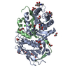 5e8mS S: Starting model for refinement C: citing same article ( |
|---|---|
| Similar structure data |
-Links
 PDBj
PDBj- Assembly
| Deposited unit | 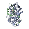
| ||||||||
|---|---|---|---|---|---|---|---|---|---|
| 1 |
| ||||||||
| Unit cell |
|
-Components
| #1: Protein | Mass: 43464.074 Da / Num. of mol.: 1 / Mutation: K307R Source method: isolated from a genetically manipulated source Source: (gene. exp.) Homo sapiens (human) / Gene: HPSE, HEP, HPA, HPA1, HPR1, HPSE1, HSE1 / Plasmid: pIEx4 / Production host:  Trichoplusia ni (cabbage looper) / References: UniProt: Q9Y251, heparanase Trichoplusia ni (cabbage looper) / References: UniProt: Q9Y251, heparanase |
|---|---|
| #2: Protein | Mass: 8273.514 Da / Num. of mol.: 1 Source method: isolated from a genetically manipulated source Source: (gene. exp.) Homo sapiens (human) / Gene: HPSE, HEP, HPA, HPA1, HPR1, HPSE1, HSE1 / Plasmid: pIEx4 / Production host: Trichoplusia ni (cabbage looper) / References: UniProt: Q9Y251, heparanase |
| #3: Chemical | ChemComp-IUV / (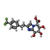  Mass: 386.322 Da / Num. of mol.: 1 / Source method: obtained synthetically / Formula: C17H17F3N2O5 / Feature type: SUBJECT OF INVESTIGATION Mass: 386.322 Da / Num. of mol.: 1 / Source method: obtained synthetically / Formula: C17H17F3N2O5 / Feature type: SUBJECT OF INVESTIGATION |
| #4: Water | ChemComp-HOH /  Mass: 18.015 Da / Num. of mol.: 13 / Source method: isolated from a natural source / Formula: H2O Mass: 18.015 Da / Num. of mol.: 13 / Source method: isolated from a natural source / Formula: H2O |
| Has ligand of interest | Y |
-Experimental details
-Experiment
| Experiment | Method: X-RAY DIFFRACTION / Number of used crystals: 1 |
|---|
- Sample preparation
| Crystal | Density Matthews: 2.28 Å3/Da / Density % sol: 45.95 % |
|---|---|
| Crystal grow | Temperature: 293 K / Method: vapor diffusion / pH: 4 Details: 12% w/v Polyethylene glycol 3,350, 100 mM Sodium malonate pH 4.0 |
-Data collection
| Diffraction | Mean temperature: 100 K / Serial crystal experiment: N |
|---|---|
| Diffraction source | Source: ROTATING ANODE / Type: RIGAKU MICROMAX-007 HF / Wavelength: 1.54184 Å |
| Detector | Type: DECTRIS PILATUS 200K / Detector: PIXEL / Date: Oct 4, 2017 |
| Radiation | Protocol: SINGLE WAVELENGTH / Monochromatic (M) / Laue (L): M / Scattering type: x-ray |
| Radiation wavelength | Wavelength: 1.54184 Å / Relative weight: 1 |
| Reflection | Resolution: 2.8→21.73 Å / Num. obs: 11532 / % possible obs: 99.4 % / Redundancy: 5.1 % / CC1/2: 0.983 / Net I/σ(I): 6.3 |
| Reflection shell | Resolution: 2.8→2.95 Å / Num. unique obs: 1661 / CC1/2: 0.722 |
- Processing
| Software |
| ||||||||||||||||||||||||||||||||||||||||||||||||||||||||||||||||||||||||||||||||||||||||||||||||||||||||||||||||||||||||||||||||||||||||||||||||||||||||||||||||||||||||||||||||||||||
|---|---|---|---|---|---|---|---|---|---|---|---|---|---|---|---|---|---|---|---|---|---|---|---|---|---|---|---|---|---|---|---|---|---|---|---|---|---|---|---|---|---|---|---|---|---|---|---|---|---|---|---|---|---|---|---|---|---|---|---|---|---|---|---|---|---|---|---|---|---|---|---|---|---|---|---|---|---|---|---|---|---|---|---|---|---|---|---|---|---|---|---|---|---|---|---|---|---|---|---|---|---|---|---|---|---|---|---|---|---|---|---|---|---|---|---|---|---|---|---|---|---|---|---|---|---|---|---|---|---|---|---|---|---|---|---|---|---|---|---|---|---|---|---|---|---|---|---|---|---|---|---|---|---|---|---|---|---|---|---|---|---|---|---|---|---|---|---|---|---|---|---|---|---|---|---|---|---|---|---|---|---|---|---|
| Refinement | Method to determine structure: MOLECULAR REPLACEMENT Starting model: 5E8M Resolution: 2.8→21.73 Å / Cor.coef. Fo:Fc: 0.944 / Cor.coef. Fo:Fc free: 0.897 / SU B: 30.68 / SU ML: 0.52 / Cross valid method: THROUGHOUT / ESU R Free: 0.467 / Stereochemistry target values: MAXIMUM LIKELIHOOD / Details: HYDROGENS HAVE BEEN ADDED IN THE RIDING POSITIONS
| ||||||||||||||||||||||||||||||||||||||||||||||||||||||||||||||||||||||||||||||||||||||||||||||||||||||||||||||||||||||||||||||||||||||||||||||||||||||||||||||||||||||||||||||||||||||
| Solvent computation | Ion probe radii: 0.8 Å / Shrinkage radii: 0.8 Å / VDW probe radii: 1.2 Å / Solvent model: MASK | ||||||||||||||||||||||||||||||||||||||||||||||||||||||||||||||||||||||||||||||||||||||||||||||||||||||||||||||||||||||||||||||||||||||||||||||||||||||||||||||||||||||||||||||||||||||
| Displacement parameters | Biso mean: 58.406 Å2
| ||||||||||||||||||||||||||||||||||||||||||||||||||||||||||||||||||||||||||||||||||||||||||||||||||||||||||||||||||||||||||||||||||||||||||||||||||||||||||||||||||||||||||||||||||||||
| Refinement step | Cycle: 1 / Resolution: 2.8→21.73 Å
| ||||||||||||||||||||||||||||||||||||||||||||||||||||||||||||||||||||||||||||||||||||||||||||||||||||||||||||||||||||||||||||||||||||||||||||||||||||||||||||||||||||||||||||||||||||||
| Refine LS restraints |
|