+ Open data
- Basic information
| Entry | Database: PDB / ID: 7tuy | ||||||||||||
|---|---|---|---|---|---|---|---|---|---|---|---|---|---|
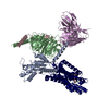
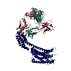
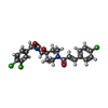
| Title | Cryo-EM structure of GSK682753A-bound EBI2/GPR183 | ||||||||||||
 Components Components |
| ||||||||||||
 Keywords Keywords | IMMUNE SYSTEM / EBI2 / GPR183 / oxysterol / GPCR / signaling / immunity / immunocyte migration | ||||||||||||
| Function / homology |  Function and homology information Function and homology informationregulation of astrocyte chemotaxis / B cell activation involved in immune response / dendritic cell homeostasis / T follicular helper cell differentiation / mature B cell differentiation involved in immune response / Class A/1 (Rhodopsin-like receptors) / T cell chemotaxis / oxysterol binding / leukocyte chemotaxis / dendritic cell chemotaxis ...regulation of astrocyte chemotaxis / B cell activation involved in immune response / dendritic cell homeostasis / T follicular helper cell differentiation / mature B cell differentiation involved in immune response / Class A/1 (Rhodopsin-like receptors) / T cell chemotaxis / oxysterol binding / leukocyte chemotaxis / dendritic cell chemotaxis / humoral immune response / positive regulation of B cell proliferation / osteoclast differentiation / G protein-coupled receptor activity / G alpha (i) signalling events / adaptive immune response / positive regulation of ERK1 and ERK2 cascade / immune response / G protein-coupled receptor signaling pathway / plasma membrane Similarity search - Function | ||||||||||||
| Biological species |  Homo sapiens (human) Homo sapiens (human)synthetic construct (others)  | ||||||||||||
| Method | ELECTRON MICROSCOPY / single particle reconstruction / cryo EM / Resolution: 2.98 Å | ||||||||||||
 Authors Authors | Chen, H. / Huang, W. / Li, X. | ||||||||||||
| Funding support |  United States, 3items United States, 3items
| ||||||||||||
 Citation Citation |  Journal: Structure / Year: 2022 Journal: Structure / Year: 2022Title: Structures of oxysterol sensor EBI2/GPR183, a key regulator of the immune response. Authors: Hongwen Chen / Weijiao Huang / Xiaochun Li /  Abstract: Oxysterols induce the migration of B-lymphocytes and dendritic cells to interfollicular regions of lymphoid tissues through binding the EBI2 (GPR183) to stimulate effective adaptive immunity and ...Oxysterols induce the migration of B-lymphocytes and dendritic cells to interfollicular regions of lymphoid tissues through binding the EBI2 (GPR183) to stimulate effective adaptive immunity and antibody production during infection. Aberrant EBI2 signaling is implicated in inflammatory bowel disease, sclerosis, and infectious disease. Here, we report the cryo-EM structures of an EBI2-G signaling complex with its endogenous agonist 7α,25-OHC and that of an inactive EBI2 bound to the inverse agonist GSK682753A. These structures reveal an agonist binding site for the oxysterol and a potential ligand entrance site exposed to the lipid bilayer. Mutations within the oxysterol binding site and the Gα interface attenuate G protein signaling and abolish oxysterol-mediated cell migration indicating that G protein signaling directly involves in the oxysterol-EBI2 pathway. Together, these findings provide new insight into how EBI2 is activated by an oxysterol ligand and will facilitate the development of therapeutic approaches that target EBI2-linked diseases. | ||||||||||||
| History |
|
- Structure visualization
| Structure viewer | Molecule:  Molmil Molmil Jmol/JSmol Jmol/JSmol |
|---|
- Downloads & links
- Download
| PDBx/mmCIF format |  7tuy.cif.gz 7tuy.cif.gz | 172.2 KB | Display |  PDBx/mmCIF format PDBx/mmCIF format |
|---|---|---|---|---|
| PDB format |  pdb7tuy.ent.gz pdb7tuy.ent.gz | 134.5 KB | Display |  PDB format PDB format |
| PDBx/mmJSON format |  7tuy.json.gz 7tuy.json.gz | Tree view |  PDBx/mmJSON format PDBx/mmJSON format | |
| Others |  Other downloads Other downloads |
-Validation report
| Summary document |  7tuy_validation.pdf.gz 7tuy_validation.pdf.gz | 818.5 KB | Display |  wwPDB validaton report wwPDB validaton report |
|---|---|---|---|---|
| Full document |  7tuy_full_validation.pdf.gz 7tuy_full_validation.pdf.gz | 827.3 KB | Display | |
| Data in XML |  7tuy_validation.xml.gz 7tuy_validation.xml.gz | 31.9 KB | Display | |
| Data in CIF |  7tuy_validation.cif.gz 7tuy_validation.cif.gz | 47.2 KB | Display | |
| Arichive directory |  https://data.pdbj.org/pub/pdb/validation_reports/tu/7tuy https://data.pdbj.org/pub/pdb/validation_reports/tu/7tuy ftp://data.pdbj.org/pub/pdb/validation_reports/tu/7tuy ftp://data.pdbj.org/pub/pdb/validation_reports/tu/7tuy | HTTPS FTP |
-Related structure data
| Related structure data |  26135MC  7tuzC M: map data used to model this data C: citing same article ( |
|---|---|
| Similar structure data | Similarity search - Function & homology  F&H Search F&H Search |
- Links
Links
- Assembly
| Deposited unit | 
|
|---|---|
| 1 |
|
- Components
| #1: Antibody | Mass: 24321.084 Da / Num. of mol.: 1 Source method: isolated from a genetically manipulated source Source: (gene. exp.)  Homo sapiens (human) Homo sapiens (human)Production host:  |
|---|---|
| #2: Antibody | Mass: 15071.431 Da / Num. of mol.: 1 Source method: isolated from a genetically manipulated source Source: (gene. exp.) synthetic construct (others) Production host:  |
| #3: Antibody | Mass: 23483.062 Da / Num. of mol.: 1 Source method: isolated from a genetically manipulated source Source: (gene. exp.)  Homo sapiens (human) Homo sapiens (human)Production host:  |
| #4: Protein | Mass: 54364.539 Da / Num. of mol.: 1 Source method: isolated from a genetically manipulated source Source: (gene. exp.)  Homo sapiens (human), (gene. exp.) Homo sapiens (human), (gene. exp.)  Gene: GPR183, EBI2 / Cell line (production host): HEK203S GnTI- / Production host:  Homo sapiens (human) / References: UniProt: P32249 Homo sapiens (human) / References: UniProt: P32249 |
| #5: Chemical | ChemComp-KKF / |
| Has ligand of interest | Y |
-Experimental details
-Experiment
| Experiment | Method: ELECTRON MICROSCOPY |
|---|---|
| EM experiment | Aggregation state: PARTICLE / 3D reconstruction method: single particle reconstruction |
- Sample preparation
| Component |
| ||||||||||||||||||||||||||||||
|---|---|---|---|---|---|---|---|---|---|---|---|---|---|---|---|---|---|---|---|---|---|---|---|---|---|---|---|---|---|---|---|
| Molecular weight | Experimental value: NO | ||||||||||||||||||||||||||||||
| Source (natural) |
| ||||||||||||||||||||||||||||||
| Source (recombinant) |
| ||||||||||||||||||||||||||||||
| Buffer solution | pH: 7.5 | ||||||||||||||||||||||||||||||
| Specimen | Embedding applied: NO / Shadowing applied: NO / Staining applied: NO / Vitrification applied: YES | ||||||||||||||||||||||||||||||
| Specimen support | Grid material: GOLD / Grid mesh size: 400 divisions/in. / Grid type: C-flat-1.2/1.3 | ||||||||||||||||||||||||||||||
| Vitrification | Cryogen name: ETHANE / Humidity: 100 % / Chamber temperature: 293 K |
- Electron microscopy imaging
| Microscopy | Model: FEI TITAN |
|---|---|
| Electron gun | Electron source:  FIELD EMISSION GUN / Accelerating voltage: 300 kV / Illumination mode: FLOOD BEAM FIELD EMISSION GUN / Accelerating voltage: 300 kV / Illumination mode: FLOOD BEAM |
| Electron lens | Mode: DIFFRACTION / Nominal defocus max: 2500 nm / Nominal defocus min: 1200 nm |
| Image recording | Electron dose: 60 e/Å2 / Film or detector model: GATAN K3 BIOQUANTUM (6k x 4k) |
- Processing
| Software | Name: PHENIX / Version: 1.16_3549: / Classification: refinement | ||||||||||||||||||||||||
|---|---|---|---|---|---|---|---|---|---|---|---|---|---|---|---|---|---|---|---|---|---|---|---|---|---|
| EM software | Name: SerialEM / Category: image acquisition | ||||||||||||||||||||||||
| CTF correction | Type: NONE | ||||||||||||||||||||||||
| 3D reconstruction | Resolution: 2.98 Å / Resolution method: FSC 0.143 CUT-OFF / Num. of particles: 222283 / Symmetry type: POINT | ||||||||||||||||||||||||
| Refine LS restraints |
|
 Movie
Movie Controller


 PDBj
PDBj


