 Movie
Movie Controller

[English] 日本語
 Yorodumi
- PDB-7ssu: Structure of EmrE-D3 mutant in complex with monobody L10 and meth... -
+ Open data

- Basic information
| Entry | Database: PDB / ID: 7ssu | ||||||
|---|---|---|---|---|---|---|---|
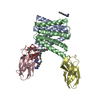
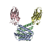
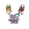
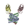
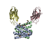
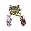
| Title | Structure of EmrE-D3 mutant in complex with monobody L10 and methyltriphenylphosphonium | ||||||
 Components Components |
| ||||||
 Keywords Keywords | TRANSPORT PROTEIN/IMMUNE SYSTEM / Small Multidrug Resistance transporters / drug efflux pump / EmrE / Membrane protein / TRANSPORT PROTEIN-IMMUNE SYSTEM complex | ||||||
| Function / homology |  Function and homology information Function and homology informationEmrE multidrug transporter complex / amino-acid betaine transmembrane transporter activity / glycine betaine transport / choline transmembrane transporter activity / choline transport / xenobiotic detoxification by transmembrane export across the plasma membrane / antiporter activity / response to osmotic stress / Antimicrobial resistance / xenobiotic transmembrane transporter activity ...EmrE multidrug transporter complex / amino-acid betaine transmembrane transporter activity / glycine betaine transport / choline transmembrane transporter activity / choline transport / xenobiotic detoxification by transmembrane export across the plasma membrane / antiporter activity / response to osmotic stress / Antimicrobial resistance / xenobiotic transmembrane transporter activity / transmembrane transporter activity / xenobiotic transport / xenobiotic metabolic process / transmembrane transport / cellular response to xenobiotic stimulus / response to xenobiotic stimulus / DNA damage response / membrane / identical protein binding / plasma membrane Similarity search - Function | ||||||
| Biological species |   Homo sapiens (human) Homo sapiens (human) | ||||||
| Method |  X-RAY DIFFRACTION / SYNCHROTRON / MOLECULAR REPLACEMENT / Resolution: 3.22 Å X-RAY DIFFRACTION / SYNCHROTRON / MOLECULAR REPLACEMENT / Resolution: 3.22 Å | ||||||
 Authors Authors | Kermani, A.A. / Stockbridge, R.B. | ||||||
| Funding support |  United States, 1items United States, 1items
| ||||||
 Citation Citation | Journal: Elife / Year: 2022 Title: Crystal structures of bacterial small multidrug resistance transporter EmrE in complex with structurally diverse substrates. Authors: Kermani, A.A. / Burata, O.E. / Koff, B.B. / Koide, A. / Koide, S. / Stockbridge, R.B. | ||||||
| History |
|
- Structure visualization
| Structure viewer | Molecule: MolmilJmol/JSmol |
|---|
- Downloads & links
-Download
| PDBx/mmCIF format | 7ssu.cif.gz | 86.9 KB | Display | PDBx/mmCIF format |
|---|---|---|---|---|
| PDB format | pdb7ssu.ent.gz | 64.7 KB | Display | PDB format |
| PDBx/mmJSON format | 7ssu.json.gz | Tree view | PDBx/mmJSON format | |
| Others |  Other downloads Other downloads |
-Validation report
| Arichive directory | https://data.pdbj.org/pub/pdb/validation_reports/ss/7ssuftp://data.pdbj.org/pub/pdb/validation_reports/ss/7ssu | HTTPS FTP |
|---|
-Related structure data
| Related structure data | 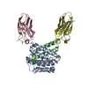 7mgxC  7mh6C  7sv9C  7svxC  7sztC  7t00C  6wk8S S: Starting model for refinement C: citing same article ( |
|---|---|
| Similar structure data |


-Links
 PDBj
PDBj

- Assembly
| Deposited unit | 
| ||||||||
|---|---|---|---|---|---|---|---|---|---|
| 1 |
| ||||||||
| Unit cell |
|
-Components
| #1: Protein | Mass: 11907.279 Da / Num. of mol.: 2 / Mutation: E25N, W31I, V34M Source method: isolated from a genetically manipulated source Source: (gene. exp.) Strain: K12 / Gene: emrE, eb, mvrC, b0543, JW0531 / Plasmid: pET-21c / Production host: #2: Antibody | Mass: 9931.934 Da / Num. of mol.: 2 Source method: isolated from a genetically manipulated source Source: (gene. exp.) Homo sapiens (human) / Production host: #3: Chemical | ChemComp-B5J / |   Mass: 277.320 Da / Num. of mol.: 1 / Source method: obtained synthetically / Formula: C19H18P / Feature type: SUBJECT OF INVESTIGATION Mass: 277.320 Da / Num. of mol.: 1 / Source method: obtained synthetically / Formula: C19H18P / Feature type: SUBJECT OF INVESTIGATIONHas ligand of interest | Y | |
|---|
-Experimental details
-Experiment
| Experiment | Method: X-RAY DIFFRACTION / Number of used crystals: 1 |
|---|
- Sample preparation
| Crystal | Density Matthews: 4.69 Å3/Da / Density % sol: 73.79 % |
|---|---|
| Crystal grow | Temperature: 294 K / Method: vapor diffusion, sitting drop / pH: 6.5 Details: 0.1 M lithium nitrate, 0.1 M ADA, pH 6.5, 33% PEG600 |
-Data collection
| Diffraction | Mean temperature: 80 K / Serial crystal experiment: N |
|---|---|
| Diffraction source | Source: SYNCHROTRON / Site: APS / Beamline: 21-ID-D / Wavelength: 0.9183 Å |
| Detector | Type: DECTRIS EIGER X 9M / Detector: PIXEL / Date: Sep 27, 2021 |
| Radiation | Protocol: SINGLE WAVELENGTH / Monochromatic (M) / Laue (L): M / Scattering type: x-ray |
| Radiation wavelength | Wavelength: 0.9183 Å / Relative weight: 1 |
| Reflection | Resolution: 3.22→70.5 Å / Num. obs: 8969 / % possible obs: 88.6 % / Redundancy: 6.6 % / CC1/2: 0.967 / Rmerge(I) obs: 0.152 / Rsym value: 0.166 / Net I/σ(I): 10.4 |
| Reflection shell | Resolution: 3.22→3.42 Å / Rmerge(I) obs: 0.656 / Num. unique obs: 448 / CC1/2: 0.861 / Rrim(I) all: 0.707 / % possible all: 13.22 |
- Processing
| Software |
| ||||||||||||||||||||||||
|---|---|---|---|---|---|---|---|---|---|---|---|---|---|---|---|---|---|---|---|---|---|---|---|---|---|
| Refinement | Method to determine structure: MOLECULAR REPLACEMENT Starting model: PDB entry 6WK8 Resolution: 3.22→58.24 Å / SU ML: 0.48 / Cross valid method: THROUGHOUT / σ(F): 1.38 / Phase error: 38.71 / Stereochemistry target values: ML
| ||||||||||||||||||||||||
| Solvent computation | Shrinkage radii: 0.9 Å / VDW probe radii: 1.11 Å / Solvent model: FLAT BULK SOLVENT MODEL | ||||||||||||||||||||||||
| Displacement parameters | Biso max: 157.42 Å2 / Biso mean: 78.5449 Å2 / Biso min: 29.3 Å2 | ||||||||||||||||||||||||
| Refinement step | Cycle: final / Resolution: 3.22→58.24 Å
| ||||||||||||||||||||||||
| LS refinement shell | Resolution: 3.22→3.34 Å /
|
