 Movie
Movie Controller

[English] 日本語
 Yorodumi
- PDB-7ssf: Light harvesting phycobiliprotein HaPE560 from the cryptophyte He... -
+ Open data

- Basic information
| Entry | Database: PDB / ID: 7ssf | ||||||||||||
|---|---|---|---|---|---|---|---|---|---|---|---|---|---|
| Title | Light harvesting phycobiliprotein HaPE560 from the cryptophyte Hemiselmis andersenii CCMP644 | ||||||||||||
 Components Components |
| ||||||||||||
 Keywords Keywords | PHOTOSYNTHESIS / phycobiliprotein / antenna / light harvesting / cryptophyte / algae / globin / CALM / CaRSP / phycoerythrin / Hemiselmis | ||||||||||||
| Function / homology |  Function and homology information Function and homology information | ||||||||||||
| Biological species |  Hemiselmis andersenii (eukaryote) Hemiselmis andersenii (eukaryote) | ||||||||||||
| Method |  X-RAY DIFFRACTION / SYNCHROTRON / MOLECULAR REPLACEMENT / Resolution: 1.45 Å X-RAY DIFFRACTION / SYNCHROTRON / MOLECULAR REPLACEMENT / Resolution: 1.45 Å | ||||||||||||
 Authors Authors | Rathbone, H.W. / Michie, K.A. / Laos, A.L. / Curmi, P.M.G. | ||||||||||||
| Funding support |  Australia, 3items Australia, 3items
| ||||||||||||
 Citation Citation | Journal: Commun Biol / Year: 2023 Title: Molecular dissection of the soluble photosynthetic antenna from the cryptophyte alga Hemiselmis andersenii Authors: Rathbone, H.W. / Curmi, P.M.G. / Michie, K.A. / Green, B.R. / Laos, A.L. / Thordarson, P. / Iranmanesh, H. #1: Journal: Acta Crystallogr D Biol Crystallogr / Year: 2012Title: Towards automated crystallographic structure refinement with phenix.refine. Authors: Afonine, P.V. / Grosse-Kunstleve, R.W. / Echols, N. / Headd, J.J. / Moriarty, N.W. / Mustyakimov, M. / Terwilliger, T.C. / Urzhumtsev, A. / Zwart, P.H. / Adams, P.D. #2: Journal: J Synchrotron Radiat / Year: 2015Title: MX1: a bending-magnet crystallography beamline serving both chemical and macromolecular crystallography communities at the Australian Synchrotron. Authors: Cowieson, N.P. / Aragao, D. / Clift, M. / Ericsson, D.J. / Gee, C. / Harrop, S.J. / Mudie, N. / Panjikar, S. / Price, J.R. / Riboldi-Tunnicliffe, A. / Williamson, R. / Caradoc-Davies, T. #3: Journal: Acta Crystallogr D Biol Crystallogr / Year: 2004Title: Coot: model-building tools for molecular graphics. Authors: Emsley, P. / Cowtan, K. #4: Journal: Acta Crystallogr D Biol Crystallogr / Year: 2011Title: Overview of the CCP4 suite and current developments. Authors: Winn, M.D. / Ballard, C.C. / Cowtan, K.D. / Dodson, E.J. / Emsley, P. / Evans, P.R. / Keegan, R.M. / Krissinel, E.B. / Leslie, A.G. / McCoy, A. / McNicholas, S.J. / Murshudov, G.N. / Pannu, ...Authors: Winn, M.D. / Ballard, C.C. / Cowtan, K.D. / Dodson, E.J. / Emsley, P. / Evans, P.R. / Keegan, R.M. / Krissinel, E.B. / Leslie, A.G. / McCoy, A. / McNicholas, S.J. / Murshudov, G.N. / Pannu, N.S. / Potterton, E.A. / Powell, H.R. / Read, R.J. / Vagin, A. / Wilson, K.S. #5: Journal: J Appl Crystallogr / Year: 2007Title: Phaser crystallographic software. Authors: McCoy, A.J. / Grosse-Kunstleve, R.W. / Adams, P.D. / Winn, M.D. / Storoni, L.C. / Read, R.J. | ||||||||||||
| History |
|
- Structure visualization
| Structure viewer | Molecule: MolmilJmol/JSmol |
|---|
- Downloads & links
-Download
| PDBx/mmCIF format | 7ssf.cif.gz | 650.2 KB | Display | PDBx/mmCIF format |
|---|---|---|---|---|
| PDB format | pdb7ssf.ent.gz | 538.8 KB | Display | PDB format |
| PDBx/mmJSON format | 7ssf.json.gz | Tree view | PDBx/mmJSON format | |
| Others |  Other downloads Other downloads |
-Validation report
| Arichive directory | https://data.pdbj.org/pub/pdb/validation_reports/ss/7ssfftp://data.pdbj.org/pub/pdb/validation_reports/ss/7ssf | HTTPS FTP |
|---|
-Related structure data
| Related structure data |  4lmxS S: Starting model for refinement |
|---|---|
| Similar structure data |
-Links
 PDBj
PDBj
- Assembly
| Deposited unit | 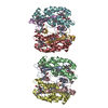
| ||||||||||||
|---|---|---|---|---|---|---|---|---|---|---|---|---|---|
| 1 | 
| ||||||||||||
| 2 | 
| ||||||||||||
| Unit cell |
| ||||||||||||
| Components on special symmetry positions |
|
-Components
-Protein , 2 types, 8 molecules ACEGBDFH
| #1: Protein | Mass: 7713.771 Da / Num. of mol.: 4 / Source method: isolated from a natural source / Source: (natural) Hemiselmis andersenii (eukaryote) / Strain: CCMP644#2: Protein | Mass: 18406.949 Da / Num. of mol.: 4 / Source method: isolated from a natural source / Source: (natural) Hemiselmis andersenii (eukaryote) / References: UniProt: U5T8W0 |
|---|
-Non-polymers , 5 types, 833 molecules 


| #3: Chemical | ChemComp-PEB /  Mass: 588.694 Da / Num. of mol.: 12 / Source method: isolated from a natural source / Formula: C33H40N4O6 / Feature type: SUBJECT OF INVESTIGATION Mass: 588.694 Da / Num. of mol.: 12 / Source method: isolated from a natural source / Formula: C33H40N4O6 / Feature type: SUBJECT OF INVESTIGATION#4: Chemical | ChemComp-AX9 /  Mass: 588.694 Da / Num. of mol.: 4 / Source method: obtained synthetically / Formula: C33H40N4O6 / Feature type: SUBJECT OF INVESTIGATION Mass: 588.694 Da / Num. of mol.: 4 / Source method: obtained synthetically / Formula: C33H40N4O6 / Feature type: SUBJECT OF INVESTIGATION#5: Chemical |  Mass: 122.143 Da / Num. of mol.: 3 / Source method: obtained synthetically / Formula: C4H12NO3 / Comment: pH buffer*YM Mass: 122.143 Da / Num. of mol.: 3 / Source method: obtained synthetically / Formula: C4H12NO3 / Comment: pH buffer*YM#6: Chemical | ChemComp-CL / |  Mass: 35.453 Da / Num. of mol.: 1 / Source method: obtained synthetically / Formula: Cl Mass: 35.453 Da / Num. of mol.: 1 / Source method: obtained synthetically / Formula: Cl#7: Water | ChemComp-HOH / | Mass: 18.015 Da / Num. of mol.: 813 / Source method: isolated from a natural source / Formula: H2O |
|---|
-Details
| Has ligand of interest | Y |
|---|---|
| Has protein modification | Y |
-Experimental details
-Experiment
| Experiment | Method: X-RAY DIFFRACTION / Number of used crystals: 1 |
|---|
- Sample preparation
| Crystal | Density Matthews: 2.53 Å3/Da / Density % sol: 51.34 % |
|---|---|
| Crystal grow | Temperature: 300 K / Method: vapor diffusion, sitting drop / pH: 6.5 / Details: TRIS HCl 0.162 M + PEGMME 5000 21.4 % (v/v) |
-Data collection
| Diffraction | Mean temperature: 100 K / Serial crystal experiment: N |
|---|---|
| Diffraction source | Source: SYNCHROTRON / Site: Australian Synchrotron / Beamline: MX1 / Wavelength: 0.9537 Å |
| Detector | Type: ADSC QUANTUM 210r / Detector: CCD / Date: Feb 23, 2019 |
| Radiation | Protocol: SINGLE WAVELENGTH / Monochromatic (M) / Laue (L): M / Scattering type: x-ray |
| Radiation wavelength | Wavelength: 0.9537 Å / Relative weight: 1 |
| Reflection | Resolution: 1.45→28.94 Å / Num. obs: 181639 / % possible obs: 99.8 % / Redundancy: 7.4 % / Biso Wilson estimate: 14.93 Å2 / CC1/2: 0.999 / Rmerge(I) obs: 0.095 / Net I/σ(I): 12.5 |
| Reflection shell | Resolution: 1.45→1.47 Å / Redundancy: 7.2 % / Rmerge(I) obs: 1.424 / Mean I/σ(I) obs: 1.4 / Num. unique obs: 8893 / CC1/2: 0.546 / % possible all: 99.3 |
- Processing
| Software |
| |||||||||||||||||||||||||||||||||||||||||||||||||||||||||||||||||||||||||||||||||||||||||||||||||||||||||||||||||||||||||||||||||||||||||||||||||||||||||||||||||||||||||||||||||||||||||||||||||||||||||||||||||||||||||
|---|---|---|---|---|---|---|---|---|---|---|---|---|---|---|---|---|---|---|---|---|---|---|---|---|---|---|---|---|---|---|---|---|---|---|---|---|---|---|---|---|---|---|---|---|---|---|---|---|---|---|---|---|---|---|---|---|---|---|---|---|---|---|---|---|---|---|---|---|---|---|---|---|---|---|---|---|---|---|---|---|---|---|---|---|---|---|---|---|---|---|---|---|---|---|---|---|---|---|---|---|---|---|---|---|---|---|---|---|---|---|---|---|---|---|---|---|---|---|---|---|---|---|---|---|---|---|---|---|---|---|---|---|---|---|---|---|---|---|---|---|---|---|---|---|---|---|---|---|---|---|---|---|---|---|---|---|---|---|---|---|---|---|---|---|---|---|---|---|---|---|---|---|---|---|---|---|---|---|---|---|---|---|---|---|---|---|---|---|---|---|---|---|---|---|---|---|---|---|---|---|---|---|---|---|---|---|---|---|---|---|---|---|---|---|---|---|---|---|
| Refinement | Method to determine structure: MOLECULAR REPLACEMENT Starting model: 4LMX Resolution: 1.45→28.94 Å / SU ML: 0.1588 / Cross valid method: FREE R-VALUE / σ(F): 1.34 / Phase error: 19.0252 Stereochemistry target values: GeoStd + Monomer Library + CDL v1.2
| |||||||||||||||||||||||||||||||||||||||||||||||||||||||||||||||||||||||||||||||||||||||||||||||||||||||||||||||||||||||||||||||||||||||||||||||||||||||||||||||||||||||||||||||||||||||||||||||||||||||||||||||||||||||||
| Solvent computation | Shrinkage radii: 0.9 Å / VDW probe radii: 1.11 Å / Solvent model: FLAT BULK SOLVENT MODEL | |||||||||||||||||||||||||||||||||||||||||||||||||||||||||||||||||||||||||||||||||||||||||||||||||||||||||||||||||||||||||||||||||||||||||||||||||||||||||||||||||||||||||||||||||||||||||||||||||||||||||||||||||||||||||
| Displacement parameters | Biso mean: 23.87 Å2 | |||||||||||||||||||||||||||||||||||||||||||||||||||||||||||||||||||||||||||||||||||||||||||||||||||||||||||||||||||||||||||||||||||||||||||||||||||||||||||||||||||||||||||||||||||||||||||||||||||||||||||||||||||||||||
| Refinement step | Cycle: LAST / Resolution: 1.45→28.94 Å
| |||||||||||||||||||||||||||||||||||||||||||||||||||||||||||||||||||||||||||||||||||||||||||||||||||||||||||||||||||||||||||||||||||||||||||||||||||||||||||||||||||||||||||||||||||||||||||||||||||||||||||||||||||||||||
| Refine LS restraints |
| |||||||||||||||||||||||||||||||||||||||||||||||||||||||||||||||||||||||||||||||||||||||||||||||||||||||||||||||||||||||||||||||||||||||||||||||||||||||||||||||||||||||||||||||||||||||||||||||||||||||||||||||||||||||||
| LS refinement shell |
|
