 Movie
Movie Controller

[English] 日本語
 Yorodumi
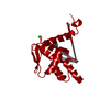
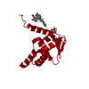
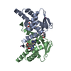
- PDB-7rv8: Crystal structure of the BCL6 BTB domain in complex with OICR-10268 -
+ Open data

- Basic information
| Entry | Database: PDB / ID: 7rv8 | ||||||
|---|---|---|---|---|---|---|---|
| Title | Crystal structure of the BCL6 BTB domain in complex with OICR-10268 | ||||||
 Components Components | Isoform 2 of B-cell lymphoma 6 protein | ||||||
 Keywords Keywords | TRANSCRIPTION / immunity / inflammatory response / transcription repressor | ||||||
| Function / homology |  Function and homology information Function and homology informationregulation of memory T cell differentiation / negative regulation of mitotic cell cycle DNA replication / intronic transcription regulatory region sequence-specific DNA binding / negative regulation of isotype switching to IgE isotypes / negative regulation of plasma cell differentiation / negative regulation of T-helper 2 cell differentiation / isotype switching to IgE isotypes / negative regulation of mast cell cytokine production / regulation of germinal center formation / negative regulation of mononuclear cell proliferation ...regulation of memory T cell differentiation / negative regulation of mitotic cell cycle DNA replication / intronic transcription regulatory region sequence-specific DNA binding / negative regulation of isotype switching to IgE isotypes / negative regulation of plasma cell differentiation / negative regulation of T-helper 2 cell differentiation / isotype switching to IgE isotypes / negative regulation of mast cell cytokine production / regulation of germinal center formation / negative regulation of mononuclear cell proliferation / plasma cell differentiation / paraspeckles / germinal center formation / pyramidal neuron differentiation / regulation of immune system process / type 2 immune response / positive regulation of regulatory T cell differentiation / T-helper 2 cell differentiation / negative regulation of B cell apoptotic process / positive regulation of cell motility / negative regulation of Rho protein signal transduction / FOXO-mediated transcription of cell death genes / negative regulation of cell-matrix adhesion / regulation of T cell proliferation / negative regulation of Notch signaling pathway / TP53 regulates transcription of several additional cell death genes whose specific roles in p53-dependent apoptosis remain uncertain / B cell proliferation / regulation of cell differentiation / negative regulation of cellular senescence / Rho protein signal transduction / regulation of immune response / erythrocyte development / heterochromatin formation / positive regulation of B cell proliferation / regulation of cytokine production / positive regulation of neuron differentiation / cell-matrix adhesion / transcription corepressor binding / cell motility / cell morphogenesis / protein localization / negative regulation of cell growth / chromatin DNA binding / DNA-binding transcription repressor activity, RNA polymerase II-specific / sequence-specific double-stranded DNA binding / regulation of cell population proliferation / regulation of inflammatory response / actin cytoskeleton organization / spermatogenesis / Interleukin-4 and Interleukin-13 signaling / DNA-binding transcription factor binding / sequence-specific DNA binding / transcription by RNA polymerase II / inflammatory response / positive regulation of apoptotic process / RNA polymerase II cis-regulatory region sequence-specific DNA binding / DNA-binding transcription factor activity / negative regulation of DNA-templated transcription / DNA damage response / chromatin binding / nucleolus / Golgi apparatus / negative regulation of transcription by RNA polymerase II / nucleoplasm / identical protein binding / nucleus / metal ion binding Similarity search - Function | ||||||
| Biological species |  Homo sapiens (human) Homo sapiens (human) | ||||||
| Method |  X-RAY DIFFRACTION / SYNCHROTRON / MOLECULAR REPLACEMENT / Resolution: 1.25 Å X-RAY DIFFRACTION / SYNCHROTRON / MOLECULAR REPLACEMENT / Resolution: 1.25 Å | ||||||
 Authors Authors | Kuntz, D.A. / Prive, G.G. | ||||||
| Funding support | 1items
| ||||||
 Citation Citation | Journal: To Be Published Title: Structure of the BCL6 BTB domain Authors: Watson, I. / Isaac, M. | ||||||
| History |
|
- Structure visualization
| Structure viewer | Molecule: MolmilJmol/JSmol |
|---|
- Downloads & links
-Download
| PDBx/mmCIF format | 7rv8.cif.gz | 121.9 KB | Display | PDBx/mmCIF format |
|---|---|---|---|---|
| PDB format | pdb7rv8.ent.gz | 94.3 KB | Display | PDB format |
| PDBx/mmJSON format | 7rv8.json.gz | Tree view | PDBx/mmJSON format | |
| Others |  Other downloads Other downloads |
-Validation report
| Summary document | 7rv8_validation.pdf.gz | 685.7 KB | Display | wwPDB validaton report |
|---|---|---|---|---|
| Full document | 7rv8_full_validation.pdf.gz | 688.3 KB | Display | |
| Data in XML | 7rv8_validation.xml.gz | 8.8 KB | Display | |
| Data in CIF | 7rv8_validation.cif.gz | 11.6 KB | Display | |
| Arichive directory | https://data.pdbj.org/pub/pdb/validation_reports/rv/7rv8ftp://data.pdbj.org/pub/pdb/validation_reports/rv/7rv8 | HTTPS FTP |
-Related structure data
| Related structure data | 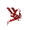 7rv3C 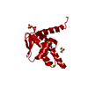 7rv4C 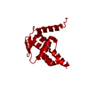 7rv5C 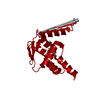 7rv6C 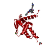 7rv7C 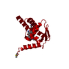 7rv9C C: citing same article ( |
|---|---|
| Similar structure data |
-Links
 PDBj
PDBj


- Assembly
| Deposited unit | 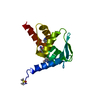
| ||||||||||||
|---|---|---|---|---|---|---|---|---|---|---|---|---|---|
| 1 | 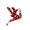
| ||||||||||||
| Unit cell |
| ||||||||||||
| Components on special symmetry positions |
|
-Components
| #1: Protein | Mass: 14559.823 Da / Num. of mol.: 1 / Mutation: C8Q,C67R,C84N Source method: isolated from a genetically manipulated source Source: (gene. exp.) Homo sapiens (human) / Gene: BCL6, BCL5, LAZ3, ZBTB27, ZNF51 / Production host:  |
|---|---|
| #2: Chemical | ChemComp-7ST / 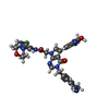  Mass: 638.076 Da / Num. of mol.: 1 / Source method: obtained synthetically / Formula: C32H28ClN9O4 / Feature type: SUBJECT OF INVESTIGATION Mass: 638.076 Da / Num. of mol.: 1 / Source method: obtained synthetically / Formula: C32H28ClN9O4 / Feature type: SUBJECT OF INVESTIGATION |
| #3: Chemical | ChemComp-DMS /   Mass: 78.133 Da / Num. of mol.: 1 / Source method: obtained synthetically / Formula: C2H6OS / Comment: DMSO, precipitant*YM Mass: 78.133 Da / Num. of mol.: 1 / Source method: obtained synthetically / Formula: C2H6OS / Comment: DMSO, precipitant*YM |
| #4: Water | ChemComp-HOH /  Mass: 18.015 Da / Num. of mol.: 103 / Source method: isolated from a natural source / Formula: H2O Mass: 18.015 Da / Num. of mol.: 103 / Source method: isolated from a natural source / Formula: H2O |
| Has ligand of interest | Y |
-Experimental details
-Experiment
| Experiment | Method: X-RAY DIFFRACTION / Number of used crystals: 1 |
|---|
- Sample preparation
| Crystal | Density Matthews: 2.03 Å3/Da / Density % sol: 39.5 % |
|---|---|
| Crystal grow | Temperature: 292 K / Method: microbatch / pH: 4.6 Details: 10% PEG6K with 0.1M MES pH 4.6, 100 mM AmSO4, 10% glycerol, 10% DMSO in Reservoir. Shifted to pH 7.4, 20% glycerol for freezing |
-Data collection
| Diffraction | Mean temperature: 100 K / Serial crystal experiment: N | ||||||||||||||||||||||||||||||
|---|---|---|---|---|---|---|---|---|---|---|---|---|---|---|---|---|---|---|---|---|---|---|---|---|---|---|---|---|---|---|---|
| Diffraction source | Source: SYNCHROTRON / Site: APS  / Beamline: 17-ID / Wavelength: 1 Å / Beamline: 17-ID / Wavelength: 1 Å | ||||||||||||||||||||||||||||||
| Detector | Type: DECTRIS PILATUS 6M / Detector: PIXEL / Date: Aug 15, 2014 | ||||||||||||||||||||||||||||||
| Radiation | Protocol: SINGLE WAVELENGTH / Monochromatic (M) / Laue (L): M / Scattering type: x-ray | ||||||||||||||||||||||||||||||
| Radiation wavelength | Wavelength: 1 Å / Relative weight: 1 | ||||||||||||||||||||||||||||||
| Reflection | Resolution: 1.25→36.28 Å / Num. obs: 30799 / % possible obs: 96.7 % / Redundancy: 3.4 % / CC1/2: 0.999 / Rmerge(I) obs: 0.034 / Rpim(I) all: 0.022 / Rrim(I) all: 0.041 / Net I/σ(I): 16.6 | ||||||||||||||||||||||||||||||
| Reflection shell | Diffraction-ID: 1
|
- Processing
| Software |
| |||||||||||||||||||||||||||||||||||||||||||||||||||||||||||||||||||||||||||
|---|---|---|---|---|---|---|---|---|---|---|---|---|---|---|---|---|---|---|---|---|---|---|---|---|---|---|---|---|---|---|---|---|---|---|---|---|---|---|---|---|---|---|---|---|---|---|---|---|---|---|---|---|---|---|---|---|---|---|---|---|---|---|---|---|---|---|---|---|---|---|---|---|---|---|---|---|
| Refinement | Method to determine structure: MOLECULAR REPLACEMENT Starting model: 1R_29 Resolution: 1.25→36.28 Å / Cor.coef. Fo:Fc: 0.978 / Cor.coef. Fo:Fc free: 0.976 / SU B: 1.719 / SU ML: 0.032 / Cross valid method: THROUGHOUT / σ(F): 0 / ESU R: 0.044 / ESU R Free: 0.042 / Stereochemistry target values: MAXIMUM LIKELIHOOD Details: HYDROGENS HAVE BEEN ADDED IN THE RIDING POSITIONS U VALUES : REFINED INDIVIDUALLY
| |||||||||||||||||||||||||||||||||||||||||||||||||||||||||||||||||||||||||||
| Solvent computation | Ion probe radii: 0.7 Å / Shrinkage radii: 0.7 Å / VDW probe radii: 1 Å / Solvent model: MASK | |||||||||||||||||||||||||||||||||||||||||||||||||||||||||||||||||||||||||||
| Displacement parameters | Biso max: 100.21 Å2 / Biso mean: 24.954 Å2 / Biso min: 11.4 Å2
| |||||||||||||||||||||||||||||||||||||||||||||||||||||||||||||||||||||||||||
| Refinement step | Cycle: final / Resolution: 1.25→36.28 Å
| |||||||||||||||||||||||||||||||||||||||||||||||||||||||||||||||||||||||||||
| Refine LS restraints |
| |||||||||||||||||||||||||||||||||||||||||||||||||||||||||||||||||||||||||||
| LS refinement shell | Resolution: 1.254→1.286 Å / Rfactor Rfree error: 0 / Total num. of bins used: 20
|
