regulation of memory T cell differentiation / negative regulation of mitotic cell cycle DNA replication / intronic transcription regulatory region sequence-specific DNA binding / negative regulation of isotype switching to IgE isotypes / negative regulation of plasma cell differentiation / negative regulation of T-helper 2 cell differentiation / isotype switching to IgE isotypes / negative regulation of mast cell cytokine production / regulation of germinal center formation / negative regulation of mononuclear cell proliferation ...regulation of memory T cell differentiation / negative regulation of mitotic cell cycle DNA replication / intronic transcription regulatory region sequence-specific DNA binding / negative regulation of isotype switching to IgE isotypes / negative regulation of plasma cell differentiation / negative regulation of T-helper 2 cell differentiation / isotype switching to IgE isotypes / negative regulation of mast cell cytokine production / regulation of germinal center formation / negative regulation of mononuclear cell proliferation / plasma cell differentiation / paraspeckles / germinal center formation / pyramidal neuron differentiation / regulation of immune system process / type 2 immune response / positive regulation of regulatory T cell differentiation / T-helper 2 cell differentiation / negative regulation of B cell apoptotic process / positive regulation of cell motility / negative regulation of Rho protein signal transduction / FOXO-mediated transcription of cell death genes / negative regulation of cell-matrix adhesion / regulation of T cell proliferation / negative regulation of Notch signaling pathway / TP53 regulates transcription of several additional cell death genes whose specific roles in p53-dependent apoptosis remain uncertain / B cell proliferation / regulation of cell differentiation / negative regulation of cellular senescence / Rho protein signal transduction / regulation of immune response / erythrocyte development / heterochromatin formation / positive regulation of B cell proliferation / regulation of cytokine production / positive regulation of neuron differentiation / cell-matrix adhesion / transcription corepressor binding / cell motility / cell morphogenesis / protein localization / negative regulation of cell growth / chromatin DNA binding / DNA-binding transcription repressor activity, RNA polymerase II-specific / sequence-specific double-stranded DNA binding / regulation of cell population proliferation / regulation of inflammatory response / actin cytoskeleton organization / spermatogenesis / Interleukin-4 and Interleukin-13 signaling / DNA-binding transcription factor binding / sequence-specific DNA binding / transcription by RNA polymerase II / inflammatory response / positive regulation of apoptotic process / RNA polymerase II cis-regulatory region sequence-specific DNA binding / DNA-binding transcription factor activity / negative regulation of DNA-templated transcription / DNA damage response / chromatin binding / nucleolus / Golgi apparatus / negative regulation of transcription by RNA polymerase II / nucleoplasm / identical protein binding / nucleus / metal ion binding Similarity search - Function
Mass: 18.015 Da / Num. of mol.: 82 / Source method: isolated from a natural source / Formula: H2O
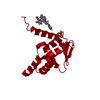
Has ligand of interest
Y
-
Experimental details
-
Experiment
Experiment
Method: X-RAY DIFFRACTION / Number of used crystals: 1
-
Sample preparation
Crystal
Density Matthews: 2.02 Å3/Da / Density % sol: 39.01 %
Crystal grow
Temperature: 292 K / Method: microbatch / pH: 6 Details: Precipitant solution was 16-20% PEG 8K in 0.1M pH 6. BisTris buffer. Crystals were soaked in 0.1M Hepes 7.4, 20% PEG 8K, 25% glycerol 0.5 mM ligand prior to passage through paratone and freezing.
-
Data collection
Diffraction
Mean temperature: 100 K / Serial crystal experiment: N
In the structure databanks used in Yorodumi, some data are registered as the other names, "COVID-19 virus" and "2019-nCoV". Here are the details of the virus and the list of structure data.
Jan 31, 2019. EMDB accession codes are about to change! (news from PDBe EMDB page)
EMDB accession codes are about to change! (news from PDBe EMDB page)
The allocation of 4 digits for EMDB accession codes will soon come to an end. Whilst these codes will remain in use, new EMDB accession codes will include an additional digit and will expand incrementally as the available range of codes is exhausted. The current 4-digit format prefixed with “EMD-” (i.e. EMD-XXXX) will advance to a 5-digit format (i.e. EMD-XXXXX), and so on. It is currently estimated that the 4-digit codes will be depleted around Spring 2019, at which point the 5-digit format will come into force.
The EM Navigator/Yorodumi systems omit the EMD- prefix.
Related info.:Q: What is EMD? / ID/Accession-code notation in Yorodumi/EM Navigator
Yorodumi is a browser for structure data from EMDB, PDB, SASBDB, etc.
This page is also the successor to EM Navigator detail page, and also detail information page/front-end page for Omokage search.
The word "yorodu" (or yorozu) is an old Japanese word meaning "ten thousand". "mi" (miru) is to see.
Related info.:EMDB / PDB / SASBDB / Comparison of 3 databanks / Yorodumi Search / Aug 31, 2016. New EM Navigator & Yorodumi / Yorodumi Papers / Jmol/JSmol / Function and homology information / Changes in new EM Navigator and Yorodumi
 Movie
Movie Controller

 Yorodumi
 Open data
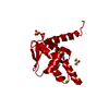
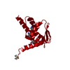
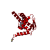
 Basic information
 Components
 Keywords
 Function and homology information
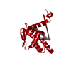
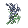
 Homo sapiens (human)
 X-RAY DIFFRACTION /
  Authors
 Canada, 1items
  Citation
 Structure visualization
 Downloads & links
 Other downloads
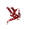


 PDBj
PDBj


 Assembly
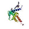


 Mass: 538.945 Da / Num. of mol.: 1 / Source method: obtained synthetically / Formula: C27H19ClN8O3 / Feature type: SUBJECT OF INVESTIGATION
 Mass: 92.094 Da / Num. of mol.: 2 / Source method: obtained synthetically / Formula: C3H8O3
 Mass: 40.078 Da / Num. of mol.: 1 / Source method: obtained synthetically / Formula: Ca
 Mass: 18.015 Da / Num. of mol.: 82 / Source method: isolated from a natural source / Formula: H2O
 Sample preparation
 / Beamline: 17-ID / Wavelength: 1 Å
 Processing