 Movie
Movie Controller


+ Open data

- Basic information
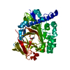
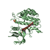
| Entry | Database: PDB / ID: 7q8z | ||||||
|---|---|---|---|---|---|---|---|
| Title | Crystal structure of TTBK2 in complex with VNG1.33 (compound 27) | ||||||
 Components Components | Tau-tubulin kinase 2 | ||||||
 Keywords Keywords | TRANSFERASE / kinase / TTBK2 / tau tubulin kinase / kinase inhibitor | ||||||
| Function / homology |  Function and homology information Function and homology informationnegative regulation of protein localization to microtubule / cerebellar granular layer development / cerebellar granule cell precursor tangential migration / positive regulation of non-motile cilium assembly / protein localization to organelle / negative regulation of microtubule depolymerization / embryonic brain development / microtubule plus-end binding / regulation of smoothened signaling pathway / ciliary transition zone ...negative regulation of protein localization to microtubule / cerebellar granular layer development / cerebellar granule cell precursor tangential migration / positive regulation of non-motile cilium assembly / protein localization to organelle / negative regulation of microtubule depolymerization / embryonic brain development / microtubule plus-end binding / regulation of smoothened signaling pathway / ciliary transition zone / neural tube development / embryonic digit morphogenesis / tau-protein kinase activity / smoothened signaling pathway / ciliary base / forebrain development / cilium assembly / kinesin binding / centriole / Anchoring of the basal body to the plasma membrane / regulation of cell migration / cerebellum development / peptidyl-serine phosphorylation / microtubule cytoskeleton organization / tau protein binding / microtubule cytoskeleton / non-specific serine/threonine protein kinase / cilium / ciliary basal body / protein serine kinase activity / protein serine/threonine kinase activity / signal transduction / extracellular space / ATP binding / nucleus / cytoplasm / cytosol Similarity search - Function | ||||||
| Biological species |  Homo sapiens (human) Homo sapiens (human) | ||||||
| Method |  X-RAY DIFFRACTION / SYNCHROTRON / MOLECULAR REPLACEMENT / Resolution: 1.57 Å X-RAY DIFFRACTION / SYNCHROTRON / MOLECULAR REPLACEMENT / Resolution: 1.57 Å | ||||||
 Authors Authors | Chaikuad, A. / Nozal, V. / Martinez, A. / Knapp, S. / Structural Genomics Consortium (SGC) | ||||||
| Funding support |  Switzerland, 1items Switzerland, 1items
| ||||||
 Citation Citation | Journal: J.Med.Chem. / Year: 2022 Title: TDP-43 Modulation by Tau-Tubulin Kinase 1 Inhibitors: A New Avenue for Future Amyotrophic Lateral Sclerosis Therapy. Authors: Nozal, V. / Martinez-Gonzalez, L. / Gomez-Almeria, M. / Gonzalo-Consuegra, C. / Santana, P. / Chaikuad, A. / Perez-Cuevas, E. / Knapp, S. / Lietha, D. / Ramirez, D. / Petralla, S. / Monti, B. ...Authors: Nozal, V. / Martinez-Gonzalez, L. / Gomez-Almeria, M. / Gonzalo-Consuegra, C. / Santana, P. / Chaikuad, A. / Perez-Cuevas, E. / Knapp, S. / Lietha, D. / Ramirez, D. / Petralla, S. / Monti, B. / Gil, C. / Martin-Requero, A. / Palomo, V. / de Lago, E. / Martinez, A. | ||||||
| History |
|
- Structure visualization
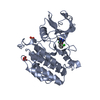
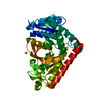
| Structure viewer | Molecule: MolmilJmol/JSmol |
|---|
- Downloads & links
-Download
| PDBx/mmCIF format | 7q8z.cif.gz | 266.6 KB | Display | PDBx/mmCIF format |
|---|---|---|---|---|
| PDB format | pdb7q8z.ent.gz | 214.8 KB | Display | PDB format |
| PDBx/mmJSON format | 7q8z.json.gz | Tree view | PDBx/mmJSON format | |
| Others |  Other downloads Other downloads |
-Validation report
| Arichive directory | https://data.pdbj.org/pub/pdb/validation_reports/q8/7q8zftp://data.pdbj.org/pub/pdb/validation_reports/q8/7q8z | HTTPS FTP |
|---|
-Related structure data
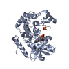
| Related structure data |  7q8vC  7q8wC  7q8yC 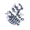 7q90C 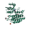 7qhwC 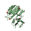 6u0kS S: Starting model for refinement C: citing same article ( |
|---|---|
| Similar structure data |


-Links
 PDBj
PDBj- Assembly
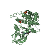
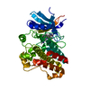
| Deposited unit | 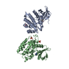
| ||||||||||||||||||
|---|---|---|---|---|---|---|---|---|---|---|---|---|---|---|---|---|---|---|---|
| 1 | 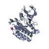
| ||||||||||||||||||
| 2 | 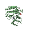
| ||||||||||||||||||
| Unit cell |
| ||||||||||||||||||
| Noncrystallographic symmetry (NCS) | NCS domain:
NCS domain segments: Component-ID: _ / Ens-ID: 1 / Beg auth comp-ID: GLN / Beg label comp-ID: GLN / End auth comp-ID: GLU / End label comp-ID: GLU / Refine code: _ / Auth seq-ID: 7 - 298 / Label seq-ID: 8 - 299
|
-Components
| #1: Protein | Mass: 34467.723 Da / Num. of mol.: 2 Source method: isolated from a genetically manipulated source Source: (gene. exp.) Homo sapiens (human) / Gene: TTBK2, KIAA0847 / Plasmid: pNIC28-Bsa4 / Production host:  References: UniProt: Q6IQ55, non-specific serine/threonine protein kinase #2: Chemical | ChemComp-PO4 /   Mass: 94.971 Da / Num. of mol.: 4 / Source method: obtained synthetically / Formula: PO4 Mass: 94.971 Da / Num. of mol.: 4 / Source method: obtained synthetically / Formula: PO4#3: Chemical | 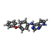  Mass: 302.330 Da / Num. of mol.: 2 / Source method: obtained synthetically / Formula: C18H14N4O / Feature type: SUBJECT OF INVESTIGATION Mass: 302.330 Da / Num. of mol.: 2 / Source method: obtained synthetically / Formula: C18H14N4O / Feature type: SUBJECT OF INVESTIGATION#4: Water | ChemComp-HOH / |  Mass: 18.015 Da / Num. of mol.: 726 / Source method: isolated from a natural source / Formula: H2O Mass: 18.015 Da / Num. of mol.: 726 / Source method: isolated from a natural source / Formula: H2OHas ligand of interest | Y | |
|---|
-Experimental details
-Experiment
| Experiment | Method: X-RAY DIFFRACTION / Number of used crystals: 1 |
|---|
- Sample preparation
| Crystal | Density Matthews: 2.81 Å3/Da / Density % sol: 56.15 % |
|---|---|
| Crystal grow | Temperature: 293.15 K / Method: vapor diffusion, sitting drop / pH: 8.5 Details: 1.6M Na/K phosphate pH 7, 5% Glycerol, 0.1M tris 8.5 |
-Data collection
| Diffraction | Mean temperature: 100 K / Serial crystal experiment: N | |||||||||||||||||||||||||||
|---|---|---|---|---|---|---|---|---|---|---|---|---|---|---|---|---|---|---|---|---|---|---|---|---|---|---|---|---|
| Diffraction source | Source: SYNCHROTRON / Site: SLS / Beamline: X06SA / Wavelength: 1 Å | |||||||||||||||||||||||||||
| Detector | Type: DECTRIS EIGER2 X 16M / Detector: PIXEL / Date: Apr 18, 2021 | |||||||||||||||||||||||||||
| Radiation | Protocol: SINGLE WAVELENGTH / Monochromatic (M) / Laue (L): M / Scattering type: x-ray | |||||||||||||||||||||||||||
| Radiation wavelength | Wavelength: 1 Å / Relative weight: 1 | |||||||||||||||||||||||||||
| Reflection | Resolution: 1.57→46.39 Å / Num. obs: 106725 / % possible obs: 98.2 % / Redundancy: 10.3 % / CC1/2: 0.998 / Rmerge(I) obs: 0.083 / Rpim(I) all: 0.029 / Rrim(I) all: 0.092 / Net I/σ(I): 15.7 | |||||||||||||||||||||||||||
| Reflection shell | Diffraction-ID: 1
|
- Processing
| Software |
| |||||||||||||||||||||||||||||||||||||||||||||||||||||||||||||||||||||||||||||||||||||||||||||||||||||||||||||||||||||||||||||||||||||||||||||||||||||||||||||||||||||||||||||||
|---|---|---|---|---|---|---|---|---|---|---|---|---|---|---|---|---|---|---|---|---|---|---|---|---|---|---|---|---|---|---|---|---|---|---|---|---|---|---|---|---|---|---|---|---|---|---|---|---|---|---|---|---|---|---|---|---|---|---|---|---|---|---|---|---|---|---|---|---|---|---|---|---|---|---|---|---|---|---|---|---|---|---|---|---|---|---|---|---|---|---|---|---|---|---|---|---|---|---|---|---|---|---|---|---|---|---|---|---|---|---|---|---|---|---|---|---|---|---|---|---|---|---|---|---|---|---|---|---|---|---|---|---|---|---|---|---|---|---|---|---|---|---|---|---|---|---|---|---|---|---|---|---|---|---|---|---|---|---|---|---|---|---|---|---|---|---|---|---|---|---|---|---|---|---|---|---|
| Refinement | Method to determine structure: MOLECULAR REPLACEMENT Starting model: 6u0k Resolution: 1.57→41.56 Å / Cor.coef. Fo:Fc: 0.965 / Cor.coef. Fo:Fc free: 0.956 / SU B: 2.831 / SU ML: 0.051 / SU R Cruickshank DPI: 0.0752 / Cross valid method: THROUGHOUT / σ(F): 0 / ESU R: 0.075 / ESU R Free: 0.077 / Stereochemistry target values: MAXIMUM LIKELIHOOD Details: HYDROGENS HAVE BEEN ADDED IN THE RIDING POSITIONS U VALUES : WITH TLS ADDED
| |||||||||||||||||||||||||||||||||||||||||||||||||||||||||||||||||||||||||||||||||||||||||||||||||||||||||||||||||||||||||||||||||||||||||||||||||||||||||||||||||||||||||||||||
| Solvent computation | Ion probe radii: 0.8 Å / Shrinkage radii: 0.8 Å / VDW probe radii: 1.2 Å / Solvent model: MASK | |||||||||||||||||||||||||||||||||||||||||||||||||||||||||||||||||||||||||||||||||||||||||||||||||||||||||||||||||||||||||||||||||||||||||||||||||||||||||||||||||||||||||||||||
| Displacement parameters | Biso max: 92.12 Å2 / Biso mean: 22.032 Å2 / Biso min: 11.1 Å2
| |||||||||||||||||||||||||||||||||||||||||||||||||||||||||||||||||||||||||||||||||||||||||||||||||||||||||||||||||||||||||||||||||||||||||||||||||||||||||||||||||||||||||||||||
| Refinement step | Cycle: final / Resolution: 1.57→41.56 Å
| |||||||||||||||||||||||||||||||||||||||||||||||||||||||||||||||||||||||||||||||||||||||||||||||||||||||||||||||||||||||||||||||||||||||||||||||||||||||||||||||||||||||||||||||
| Refine LS restraints |
| |||||||||||||||||||||||||||||||||||||||||||||||||||||||||||||||||||||||||||||||||||||||||||||||||||||||||||||||||||||||||||||||||||||||||||||||||||||||||||||||||||||||||||||||
| Refine LS restraints NCS | Ens-ID: 1 / Number: 9415 / Refine-ID: X-RAY DIFFRACTION / Type: interatomic distance / Rms dev position: 0.12 Å / Weight position: 0.05
| |||||||||||||||||||||||||||||||||||||||||||||||||||||||||||||||||||||||||||||||||||||||||||||||||||||||||||||||||||||||||||||||||||||||||||||||||||||||||||||||||||||||||||||||
| LS refinement shell | Resolution: 1.57→1.611 Å / Rfactor Rfree error: 0 / Total num. of bins used: 20
| |||||||||||||||||||||||||||||||||||||||||||||||||||||||||||||||||||||||||||||||||||||||||||||||||||||||||||||||||||||||||||||||||||||||||||||||||||||||||||||||||||||||||||||||
| Refinement TLS params. | Method: refined / Refine-ID: X-RAY DIFFRACTION
| |||||||||||||||||||||||||||||||||||||||||||||||||||||||||||||||||||||||||||||||||||||||||||||||||||||||||||||||||||||||||||||||||||||||||||||||||||||||||||||||||||||||||||||||
| Refinement TLS group |
|
