 Movie
Movie Controller


+ Open data

- Basic information
| Entry | Database: PDB / ID: 7qhw | |||||||||
|---|---|---|---|---|---|---|---|---|---|---|
| Title | TTBK1 kinase domain in complex with inhibitor 29 | |||||||||
 Components Components | Tau-tubulin kinase 1 | |||||||||
 Keywords Keywords | TRANSFERASE / kinase / inhibitor | |||||||||
| Function / homology |  Function and homology information Function and homology informationpositive regulation of astrocyte activation / positive regulation of microglial cell activation / microtubule associated complex / tau-protein kinase activity / positive regulation of protein polymerization / peptidyl-threonine phosphorylation / substantia nigra development / peptidyl-tyrosine phosphorylation / peptidyl-serine phosphorylation / tau protein binding ...positive regulation of astrocyte activation / positive regulation of microglial cell activation / microtubule associated complex / tau-protein kinase activity / positive regulation of protein polymerization / peptidyl-threonine phosphorylation / substantia nigra development / peptidyl-tyrosine phosphorylation / peptidyl-serine phosphorylation / tau protein binding / protein tyrosine kinase activity / learning or memory / non-specific serine/threonine protein kinase / negative regulation of gene expression / protein serine kinase activity / protein serine/threonine kinase activity / neuronal cell body / positive regulation of gene expression / perinuclear region of cytoplasm / signal transduction / nucleoplasm / ATP binding / nucleus / cytoplasm / cytosol Similarity search - Function | |||||||||
| Biological species |  Homo sapiens (human) Homo sapiens (human) | |||||||||
| Method |  X-RAY DIFFRACTION / SYNCHROTRON / MOLECULAR REPLACEMENT / Resolution: 2.8 Å X-RAY DIFFRACTION / SYNCHROTRON / MOLECULAR REPLACEMENT / Resolution: 2.8 Å | |||||||||
 Authors Authors | Nozal, V. / Liehta, D. | |||||||||
| Funding support |  Spain, 2items Spain, 2items
| |||||||||
 Citation Citation | Journal: J.Med.Chem. / Year: 2022 Title: TDP-43 Modulation by Tau-Tubulin Kinase 1 Inhibitors: A New Avenue for Future Amyotrophic Lateral Sclerosis Therapy. Authors: Nozal, V. / Martinez-Gonzalez, L. / Gomez-Almeria, M. / Gonzalo-Consuegra, C. / Santana, P. / Chaikuad, A. / Perez-Cuevas, E. / Knapp, S. / Lietha, D. / Ramirez, D. / Petralla, S. / Monti, B. ...Authors: Nozal, V. / Martinez-Gonzalez, L. / Gomez-Almeria, M. / Gonzalo-Consuegra, C. / Santana, P. / Chaikuad, A. / Perez-Cuevas, E. / Knapp, S. / Lietha, D. / Ramirez, D. / Petralla, S. / Monti, B. / Gil, C. / Martin-Requero, A. / Palomo, V. / de Lago, E. / Martinez, A. | |||||||||
| History |
|
- Structure visualization
| Structure viewer | Molecule: MolmilJmol/JSmol |
|---|
- Downloads & links
-Download
| PDBx/mmCIF format | 7qhw.cif.gz | 192 KB | Display | PDBx/mmCIF format |
|---|---|---|---|---|
| PDB format | pdb7qhw.ent.gz | Display | PDB format | |
| PDBx/mmJSON format | 7qhw.json.gz | Tree view | PDBx/mmJSON format | |
| Others |  Other downloads Other downloads |
-Validation report
| Arichive directory | https://data.pdbj.org/pub/pdb/validation_reports/qh/7qhwftp://data.pdbj.org/pub/pdb/validation_reports/qh/7qhw | HTTPS FTP |
|---|
-Related structure data
| Related structure data |  7q8vC  7q8wC 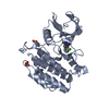 7q8yC  7q8zC 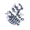 7q90C  4btmS S: Starting model for refinement C: citing same article ( |
|---|---|
| Similar structure data |
-Links
 PDBj
PDBj- Assembly
| Deposited unit | 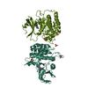
| ||||||||
|---|---|---|---|---|---|---|---|---|---|
| 1 | 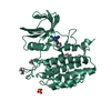
| ||||||||
| 2 | 
| ||||||||
| Unit cell |
|
-Components
| #1: Protein | Mass: 33965.352 Da / Num. of mol.: 2 Source method: isolated from a genetically manipulated source Source: (gene. exp.) Homo sapiens (human) / Gene: TTBK1, BDTK, KIAA1855 / Production host:  References: UniProt: Q5TCY1, non-specific serine/threonine protein kinase #2: Chemical | 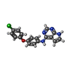  Mass: 336.775 Da / Num. of mol.: 2 / Source method: obtained synthetically / Formula: C18H13ClN4O / Feature type: SUBJECT OF INVESTIGATION Mass: 336.775 Da / Num. of mol.: 2 / Source method: obtained synthetically / Formula: C18H13ClN4O / Feature type: SUBJECT OF INVESTIGATION#3: Chemical | ChemComp-SO4 /   Mass: 96.063 Da / Num. of mol.: 4 / Source method: obtained synthetically / Formula: SO4 Mass: 96.063 Da / Num. of mol.: 4 / Source method: obtained synthetically / Formula: SO4#4: Chemical |   Mass: 92.094 Da / Num. of mol.: 3 / Source method: obtained synthetically / Formula: C3H8O3 Mass: 92.094 Da / Num. of mol.: 3 / Source method: obtained synthetically / Formula: C3H8O3#5: Water | ChemComp-HOH / |  Mass: 18.015 Da / Num. of mol.: 73 / Source method: isolated from a natural source / Formula: H2O Mass: 18.015 Da / Num. of mol.: 73 / Source method: isolated from a natural source / Formula: H2OHas ligand of interest | Y | |
|---|
-Experimental details
-Experiment
| Experiment | Method: X-RAY DIFFRACTION / Number of used crystals: 1 |
|---|
- Sample preparation
| Crystal | Density Matthews: 5.54 Å3/Da / Density % sol: 77.78 % |
|---|---|
| Crystal grow | Temperature: 293 K / Method: vapor diffusion, hanging drop / pH: 5.6 Details: 27% PEG 4000, 200mM NH4SO4, 100 mM Na Citrate pH5.6 and 10 mM TCEP |
-Data collection
| Diffraction | Mean temperature: 100 K / Serial crystal experiment: N |
|---|---|
| Diffraction source | Source: SYNCHROTRON / Site: ALBA / Beamline: XALOC / Wavelength: 0.979 Å |
| Detector | Type: DECTRIS PILATUS 6M / Detector: PIXEL / Date: Apr 21, 2021 |
| Radiation | Protocol: SINGLE WAVELENGTH / Monochromatic (M) / Laue (L): M / Scattering type: x-ray |
| Radiation wavelength | Wavelength: 0.979 Å / Relative weight: 1 |
| Reflection | Resolution: 2.8→48.69 Å / Num. obs: 36393 / % possible obs: 99.5 % / Redundancy: 3.4 % / CC1/2: 0.992 / Net I/σ(I): 5 |
| Reflection shell | Resolution: 2.8→2.873 Å / Num. unique obs: 2552 / CC1/2: 0.441 / % possible all: 99.78 |
- Processing
| Software |
| |||||||||||||||||||||||||||||||||||||||||||||||||||||||||||||||||||||||||||||||||||||||||||||||||||||||||||||||||||||||||||||||||||||||||||||||||||
|---|---|---|---|---|---|---|---|---|---|---|---|---|---|---|---|---|---|---|---|---|---|---|---|---|---|---|---|---|---|---|---|---|---|---|---|---|---|---|---|---|---|---|---|---|---|---|---|---|---|---|---|---|---|---|---|---|---|---|---|---|---|---|---|---|---|---|---|---|---|---|---|---|---|---|---|---|---|---|---|---|---|---|---|---|---|---|---|---|---|---|---|---|---|---|---|---|---|---|---|---|---|---|---|---|---|---|---|---|---|---|---|---|---|---|---|---|---|---|---|---|---|---|---|---|---|---|---|---|---|---|---|---|---|---|---|---|---|---|---|---|---|---|---|---|---|---|---|---|
| Refinement | Method to determine structure: MOLECULAR REPLACEMENT Starting model: 4BTM Resolution: 2.8→48.686 Å / Cor.coef. Fo:Fc: 0.947 / Cor.coef. Fo:Fc free: 0.932 / SU B: 14.942 / SU ML: 0.216 / Cross valid method: FREE R-VALUE / ESU R: 0.329 / ESU R Free: 0.246 Details: Hydrogens have been used if present in the input file
| |||||||||||||||||||||||||||||||||||||||||||||||||||||||||||||||||||||||||||||||||||||||||||||||||||||||||||||||||||||||||||||||||||||||||||||||||||
| Solvent computation | Ion probe radii: 0.8 Å / Shrinkage radii: 0.8 Å / VDW probe radii: 1.2 Å / Solvent model: MASK BULK SOLVENT | |||||||||||||||||||||||||||||||||||||||||||||||||||||||||||||||||||||||||||||||||||||||||||||||||||||||||||||||||||||||||||||||||||||||||||||||||||
| Displacement parameters | Biso mean: 68.455 Å2
| |||||||||||||||||||||||||||||||||||||||||||||||||||||||||||||||||||||||||||||||||||||||||||||||||||||||||||||||||||||||||||||||||||||||||||||||||||
| Refinement step | Cycle: LAST / Resolution: 2.8→48.686 Å
| |||||||||||||||||||||||||||||||||||||||||||||||||||||||||||||||||||||||||||||||||||||||||||||||||||||||||||||||||||||||||||||||||||||||||||||||||||
| Refine LS restraints |
| |||||||||||||||||||||||||||||||||||||||||||||||||||||||||||||||||||||||||||||||||||||||||||||||||||||||||||||||||||||||||||||||||||||||||||||||||||
| LS refinement shell |
| |||||||||||||||||||||||||||||||||||||||||||||||||||||||||||||||||||||||||||||||||||||||||||||||||||||||||||||||||||||||||||||||||||||||||||||||||||
| Refinement TLS params. | Method: refined / Refine-ID: X-RAY DIFFRACTION
| |||||||||||||||||||||||||||||||||||||||||||||||||||||||||||||||||||||||||||||||||||||||||||||||||||||||||||||||||||||||||||||||||||||||||||||||||||
| Refinement TLS group |
|
