 Movie
Movie Controller

[English] 日本語
 Yorodumi
- PDB-7p1m: Galectin-8 N-terminal carbohydrate recognition domain in complex ... -
+ Open data

- Basic information
| Entry | Database: PDB / ID: 7p1m | ||||||
|---|---|---|---|---|---|---|---|
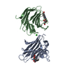
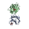
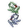
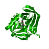
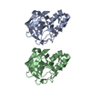
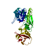
| Title | Galectin-8 N-terminal carbohydrate recognition domain in complex with benzimidazole D-galactal ligand | ||||||
 Components Components | Galectin-8 | ||||||
 Keywords Keywords | SUGAR BINDING PROTEIN / Inhibitor / complex / Galectin / Lectin / D-galactal | ||||||
| Function / homology |  Function and homology information Function and homology informationlymphatic endothelial cell migration / xenophagy / plasma cell differentiation / T cell costimulation / cellular response to virus / integrin binding / carbohydrate binding / cytoplasmic vesicle / : / membrane ...lymphatic endothelial cell migration / xenophagy / plasma cell differentiation / T cell costimulation / cellular response to virus / integrin binding / carbohydrate binding / cytoplasmic vesicle / : / membrane / cytoplasm / cytosol Similarity search - Function | ||||||
| Biological species |  Homo sapiens (human) Homo sapiens (human) | ||||||
| Method |  X-RAY DIFFRACTION / SYNCHROTRON / MOLECULAR REPLACEMENT / Resolution: 1.52 Å X-RAY DIFFRACTION / SYNCHROTRON / MOLECULAR REPLACEMENT / Resolution: 1.52 Å | ||||||
 Authors Authors | Hassan, M. / Hakansson, M. / Nilsson, J.U. / Kovacic, R. | ||||||
| Funding support |  Sweden, 1items Sweden, 1items
| ||||||
 Citation Citation | Journal: Acs Med.Chem.Lett. / Year: 2021 Title: Structure-Guided Design of d-Galactal Derivatives with High Affinity and Selectivity for the Galectin-8 N-Terminal Domain. Authors: Hassan, M. / Baussiere, F. / Guzelj, S. / Sundin, A.P. / Hakansson, M. / Kovacic, R. / Leffler, H. / Tomasic, T. / Anderluh, M. / Jakopin, Z. / Nilsson, U.J. | ||||||
| History |
|
- Structure visualization
| Structure viewer | Molecule: MolmilJmol/JSmol |
|---|
- Downloads & links
-Download
| PDBx/mmCIF format | 7p1m.cif.gz | 154.8 KB | Display | PDBx/mmCIF format |
|---|---|---|---|---|
| PDB format | pdb7p1m.ent.gz | 121.5 KB | Display | PDB format |
| PDBx/mmJSON format | 7p1m.json.gz | Tree view | PDBx/mmJSON format | |
| Others |  Other downloads Other downloads |
-Validation report
| Arichive directory | https://data.pdbj.org/pub/pdb/validation_reports/p1/7p1mftp://data.pdbj.org/pub/pdb/validation_reports/p1/7p1m | HTTPS FTP |
|---|
-Related structure data
| Related structure data | 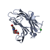 7p11C  5gzdS S: Starting model for refinement C: citing same article ( |
|---|---|
| Similar structure data |


-Links
 PDBj
PDBj- Assembly
| Deposited unit | 
| |||||||||||||||||||||||||||
|---|---|---|---|---|---|---|---|---|---|---|---|---|---|---|---|---|---|---|---|---|---|---|---|---|---|---|---|---|
| 1 |
| |||||||||||||||||||||||||||
| Unit cell |
| |||||||||||||||||||||||||||
| Noncrystallographic symmetry (NCS) | NCS domain:
NCS domain segments:
|
-Components
| #1: Protein | Mass: 17081.572 Da / Num. of mol.: 2 Source method: isolated from a genetically manipulated source Source: (gene. exp.) Homo sapiens (human) / Gene: LGALS8 / Production host:  #2: Chemical |   Mass: 334.324 Da / Num. of mol.: 2 / Source method: obtained synthetically / Formula: C16H18N2O6 / Feature type: SUBJECT OF INVESTIGATION Mass: 334.324 Da / Num. of mol.: 2 / Source method: obtained synthetically / Formula: C16H18N2O6 / Feature type: SUBJECT OF INVESTIGATION#3: Chemical | ChemComp-CL / |   Mass: 35.453 Da / Num. of mol.: 1 / Source method: obtained synthetically / Formula: Cl Mass: 35.453 Da / Num. of mol.: 1 / Source method: obtained synthetically / Formula: Cl#4: Water | ChemComp-HOH / |  Mass: 18.015 Da / Num. of mol.: 283 / Source method: isolated from a natural source / Formula: H2O Mass: 18.015 Da / Num. of mol.: 283 / Source method: isolated from a natural source / Formula: H2OHas ligand of interest | Y | |
|---|
-Experimental details
-Experiment
| Experiment | Method: X-RAY DIFFRACTION / Number of used crystals: 1 |
|---|
- Sample preparation
| Crystal | Density Matthews: 2.01 Å3/Da / Density % sol: 38.85 % |
|---|---|
| Crystal grow | Temperature: 293.5 K / Method: liquid diffusion / pH: 8 Details: Buffer (10mM Tris/HCl pH 8.0, 150 mM NaCl, 10 mm lactose and 1 mM TCEP) mixed with reservoir 25% (w/v) PEG 2000 monomethylethyer (MME), a cryo solution (10 mM Tris/HCl pH 8.0, 50 mM NaCl, 10 ...Details: Buffer (10mM Tris/HCl pH 8.0, 150 mM NaCl, 10 mm lactose and 1 mM TCEP) mixed with reservoir 25% (w/v) PEG 2000 monomethylethyer (MME), a cryo solution (10 mM Tris/HCl pH 8.0, 50 mM NaCl, 10 mM lactose, 25% (w/v) PEG 2000 MME, 20% ethylene glycol (EG) and 1 mM TCEP) and flash-frozen in liquid nitrogen. A co-crystal with lactose, grown from a seeded drop with 24% (w/v) PEG 2000 MME in the reservoir, was used to soak in compound 1 by transferring crystals in three steps to different soaking drops. First to a 2 ul drop with glycerol ( 20% (v/v) glycerol, 25% (w/v PEG 2000 MME, 10 mM Tris/HCl pH 8.0, 50 mM NaCl and 1 mM TCEP) and secondly to a 2 ul drop with 10 mM compound 1 (10 mM Tris/HCl pH 8.0, 50 mM NaCl, 5 mM compound 1, 25% (w/v) PEG 2000 MME and 1 mM TCEP) and thirdly to a second drop with 5 mM compound 1 (same composition as before). |
-Data collection
| Diffraction | Mean temperature: 293.5 K / Serial crystal experiment: N |
|---|---|
| Diffraction source | Source: SYNCHROTRON / Site: Diamond  / Beamline: I03 / Wavelength: 0.97935 Å / Beamline: I03 / Wavelength: 0.97935 Å |
| Detector | Type: DECTRIS EIGER X 16M / Detector: PIXEL / Date: Oct 22, 2019 |
| Radiation | Protocol: SINGLE WAVELENGTH / Monochromatic (M) / Laue (L): M / Scattering type: x-ray |
| Radiation wavelength | Wavelength: 0.97935 Å / Relative weight: 1 |
| Reflection | Resolution: 1.52→49.91 Å / Num. obs: 3108 / % possible obs: 99.94 % / Redundancy: 6.6 % / Rmerge(I) obs: 0.039 / Net I/σ(I): 19.1 |
| Reflection shell | Resolution: 1.52→1.56 Å / Rmerge(I) obs: 1.61 / Num. unique obs: 0 |
- Processing
| Software |
| ||||||||||||||||||||||||||||||||||||||||||||||||||||||||||||||||||||||||||||||||||||||||||||||||||||||||||||||||||||||||||||||||||||||||||||||||||||||||||||||||||||||||||||||||||||||
|---|---|---|---|---|---|---|---|---|---|---|---|---|---|---|---|---|---|---|---|---|---|---|---|---|---|---|---|---|---|---|---|---|---|---|---|---|---|---|---|---|---|---|---|---|---|---|---|---|---|---|---|---|---|---|---|---|---|---|---|---|---|---|---|---|---|---|---|---|---|---|---|---|---|---|---|---|---|---|---|---|---|---|---|---|---|---|---|---|---|---|---|---|---|---|---|---|---|---|---|---|---|---|---|---|---|---|---|---|---|---|---|---|---|---|---|---|---|---|---|---|---|---|---|---|---|---|---|---|---|---|---|---|---|---|---|---|---|---|---|---|---|---|---|---|---|---|---|---|---|---|---|---|---|---|---|---|---|---|---|---|---|---|---|---|---|---|---|---|---|---|---|---|---|---|---|---|---|---|---|---|---|---|---|
| Refinement | Method to determine structure: MOLECULAR REPLACEMENT Starting model: 5GZD Resolution: 1.52→49.91 Å / Cor.coef. Fo:Fc: 0.981 / Cor.coef. Fo:Fc free: 0.962 / SU B: 6.574 / SU ML: 0.094 / Cross valid method: THROUGHOUT / ESU R: 0.091 / ESU R Free: 0.083 / Stereochemistry target values: MAXIMUM LIKELIHOOD
| ||||||||||||||||||||||||||||||||||||||||||||||||||||||||||||||||||||||||||||||||||||||||||||||||||||||||||||||||||||||||||||||||||||||||||||||||||||||||||||||||||||||||||||||||||||||
| Solvent computation | Ion probe radii: 0.8 Å / Shrinkage radii: 0.8 Å / VDW probe radii: 1.2 Å / Solvent model: MASK | ||||||||||||||||||||||||||||||||||||||||||||||||||||||||||||||||||||||||||||||||||||||||||||||||||||||||||||||||||||||||||||||||||||||||||||||||||||||||||||||||||||||||||||||||||||||
| Displacement parameters | Biso mean: 31.956 Å2
| ||||||||||||||||||||||||||||||||||||||||||||||||||||||||||||||||||||||||||||||||||||||||||||||||||||||||||||||||||||||||||||||||||||||||||||||||||||||||||||||||||||||||||||||||||||||
| Refinement step | Cycle: 1 / Resolution: 1.52→49.91 Å
| ||||||||||||||||||||||||||||||||||||||||||||||||||||||||||||||||||||||||||||||||||||||||||||||||||||||||||||||||||||||||||||||||||||||||||||||||||||||||||||||||||||||||||||||||||||||
| Refine LS restraints |
|