 Movie
Movie Controller


+ Open data

- Basic information
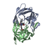
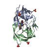
| Entry | Database: PDB / ID: 7leh | ||||||
|---|---|---|---|---|---|---|---|
| Title | HIV-1 Protease WT (NL4-3) in Complex with PU9 (LR2-80) | ||||||
 Components Components | Protease | ||||||
 Keywords Keywords | HYDROLASE/HYDROLASE INHIBITOR / HIV / NL4-3 PROTEASE / PROTEASE INHIBITOR / HYDROLASE-HYDROLASE INHIBITOR COMPLEX | ||||||
| Function / homology |  Function and homology information Function and homology informationhost multivesicular body / aspartic-type endopeptidase activity / virion membrane / proteolysis Similarity search - Function | ||||||
| Biological species |   Human immunodeficiency virus 1 Human immunodeficiency virus 1 | ||||||
| Method |  X-RAY DIFFRACTION / MOLECULAR REPLACEMENT / Resolution: 2 Å X-RAY DIFFRACTION / MOLECULAR REPLACEMENT / Resolution: 2 Å | ||||||
 Authors Authors | Lockbaum, G.J. / Rusere, L.N. / Henes, M. / Kosovrasti, K. / Lee, S.K. / Spielvogel, E. / Nalivaika, E.A. / Swanstrom, R. / KurtYilmaz, N. / Schiffer, C.A. / Ali, A. | ||||||
| Funding support |  United States, 1items United States, 1items
| ||||||
 Citation Citation | Journal: To Be Published Title: HIV-1 Protease Inhibitors with a P1 Phosphonate Modification Maintain Potency against Drug Resistant Variants by Increased van der Waals Contacts with Flaps Residues Authors: Lockbaum, G.J. / Rusere, L.N. / Henes, M. / Kosovrasti, K. / Lee, S.K. / Spielvogel, E. / Nalivaika, E.A. / Swanstrom, R. / KurtYilmaz, N. / Schiffer, C.A. / Ali, A. | ||||||
| History |
|
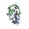
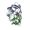
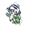
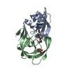
- Structure visualization
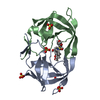
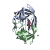
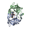
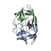
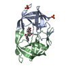
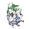
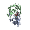
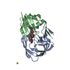
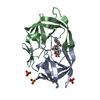
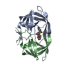
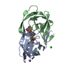
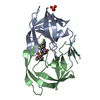
| Structure viewer | Molecule: MolmilJmol/JSmol |
|---|
- Downloads & links
-Download
| PDBx/mmCIF format | 7leh.cif.gz | 90.5 KB | Display | PDBx/mmCIF format |
|---|---|---|---|---|
| PDB format | pdb7leh.ent.gz | 68.9 KB | Display | PDB format |
| PDBx/mmJSON format | 7leh.json.gz | Tree view | PDBx/mmJSON format | |
| Others |  Other downloads Other downloads |
-Validation report
| Arichive directory | https://data.pdbj.org/pub/pdb/validation_reports/le/7lehftp://data.pdbj.org/pub/pdb/validation_reports/le/7leh | HTTPS FTP |
|---|
-Related structure data
| Related structure data | 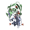 7ldyC 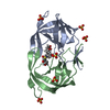 7ldzC 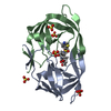 7le0C  7le1C 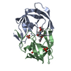 7le2C  7le3C 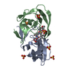 7le4C  7le5C  7le6C  7le7C  7le8C  7le9C  7leaC  7lebC  7lecC  7ledC  7leeC  7lefC  7legC 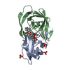 7leiC  6dgxS S: Starting model for refinement C: citing same article ( |
|---|---|
| Similar structure data |


-Links
 PDBj
PDBj
- Assembly
| Deposited unit | 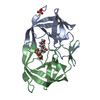
| ||||||||
|---|---|---|---|---|---|---|---|---|---|
| 1 |
| ||||||||
| Unit cell |
|
-Components
| #1: Protein | Mass: 10831.833 Da / Num. of mol.: 2 Source method: isolated from a genetically manipulated source Source: (gene. exp.) Human immunodeficiency virus 1 / Gene: pol / Plasmid: pXC35 / Production host:  #2: Chemical | ChemComp-XW4 / | 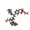  Mass: 770.824 Da / Num. of mol.: 1 / Source method: obtained synthetically / Formula: C35H51N2O13PS / Feature type: SUBJECT OF INVESTIGATION Mass: 770.824 Da / Num. of mol.: 1 / Source method: obtained synthetically / Formula: C35H51N2O13PS / Feature type: SUBJECT OF INVESTIGATION#3: Chemical | ChemComp-SO4 / |   Mass: 96.063 Da / Num. of mol.: 1 / Source method: isolated from a natural source / Formula: SO4 Mass: 96.063 Da / Num. of mol.: 1 / Source method: isolated from a natural source / Formula: SO4#4: Water | ChemComp-HOH / |  Mass: 18.015 Da / Num. of mol.: 155 / Source method: isolated from a natural source / Formula: H2O Mass: 18.015 Da / Num. of mol.: 155 / Source method: isolated from a natural source / Formula: H2OHas ligand of interest | Y | |
|---|
-Experimental details
-Experiment
| Experiment | Method: X-RAY DIFFRACTION / Number of used crystals: 1 |
|---|
- Sample preparation
| Crystal | Density Matthews: 2.14 Å3/Da / Density % sol: 42.64 % |
|---|---|
| Crystal grow | Temperature: 293 K / Method: vapor diffusion, hanging drop Details: 23-24% (w/v) Ammonium Sulfate, 0.1M Bis-Tris-Methane-HCl Buffer pH 5.5 |
-Data collection
| Diffraction | Mean temperature: 100 K / Serial crystal experiment: N |
|---|---|
| Diffraction source | Source: ROTATING ANODE / Type: RIGAKU MICROMAX-007 HF / Wavelength: 1.54178 Å |
| Detector | Type: RIGAKU SATURN 944 / Detector: CCD / Date: Jul 5, 2017 |
| Radiation | Protocol: SINGLE WAVELENGTH / Monochromatic (M) / Laue (L): M / Scattering type: x-ray |
| Radiation wavelength | Wavelength: 1.54178 Å / Relative weight: 1 |
| Reflection | Resolution: 1.996→24.2 Å / Num. obs: 12640 / % possible obs: 95.9 % / Redundancy: 7.1 % / Biso Wilson estimate: 24.35 Å2 / Rmerge(I) obs: 0.084 / Net I/σ(I): 22.6 |
| Reflection shell | Resolution: 1.996→2.02 Å / Redundancy: 6.4 % / Rmerge(I) obs: 0.399 / Mean I/σ(I) obs: 4.6 / Num. unique obs: 602 / % possible all: 93.2 |
- Processing
| Software |
| |||||||||||||||||||||||||||||||||||
|---|---|---|---|---|---|---|---|---|---|---|---|---|---|---|---|---|---|---|---|---|---|---|---|---|---|---|---|---|---|---|---|---|---|---|---|---|
| Refinement | Method to determine structure: MOLECULAR REPLACEMENT Starting model: 6DGX Resolution: 2→24.17 Å / SU ML: 0.24 / Cross valid method: THROUGHOUT / σ(F): 1.34 / Phase error: 24.99 / Stereochemistry target values: ML
| |||||||||||||||||||||||||||||||||||
| Solvent computation | Shrinkage radii: 0.9 Å / VDW probe radii: 1.11 Å / Solvent model: FLAT BULK SOLVENT MODEL | |||||||||||||||||||||||||||||||||||
| Displacement parameters | Biso max: 148.69 Å2 / Biso mean: 31.8109 Å2 / Biso min: 11.82 Å2 | |||||||||||||||||||||||||||||||||||
| Refinement step | Cycle: final / Resolution: 2→24.17 Å
| |||||||||||||||||||||||||||||||||||
| LS refinement shell | Refine-ID: X-RAY DIFFRACTION / Rfactor Rfree error: 0 / Total num. of bins used: 4
|
