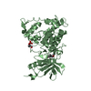
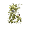
regulation of amyloid-beta formation / regulation of neurofibrillary tangle assembly / histone H3T45 kinase activity / negative regulation of heterochromatin formation / dual-specificity kinase / splicing factor binding / [RNA-polymerase]-subunit kinase / tau-protein kinase activity / regulation of alternative mRNA splicing, via spliceosome / negative regulation of microtubule polymerization ...regulation of amyloid-beta formation / regulation of neurofibrillary tangle assembly / histone H3T45 kinase activity / negative regulation of heterochromatin formation / dual-specificity kinase / splicing factor binding / [RNA-polymerase]-subunit kinase / tau-protein kinase activity / regulation of alternative mRNA splicing, via spliceosome / negative regulation of microtubule polymerization / negative regulation of DNA damage response, signal transduction by p53 class mediator / negative regulation of mRNA splicing, via spliceosome / G0 and Early G1 / cytoskeletal protein binding / peptidyl-tyrosine phosphorylation / RNA polymerase II CTD heptapeptide repeat kinase activity / protein serine/threonine/tyrosine kinase activity / positive regulation of RNA splicing / non-membrane spanning protein tyrosine kinase activity / tubulin binding / circadian rhythm / tau protein binding / protein autophosphorylation / nervous system development / actin binding / protein tyrosine kinase activity / protein phosphorylation / protein kinase activity / transcription coactivator activity / nuclear speck / ribonucleoprotein complex / protein serine kinase activity / axon / protein serine/threonine kinase activity / centrosome / dendrite / positive regulation of DNA-templated transcription / nucleoplasm / ATP binding / identical protein binding / nucleus / cytoplasm / cytosol Similarity search - Function
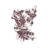
Dual specificity tyrosine-phosphorylation-regulated kinase 1A/1B, catalytic domain / : / Serine/threonine-protein kinase, active site / Serine/Threonine protein kinases active-site signature. / Protein kinase domain / Serine/Threonine protein kinases, catalytic domain / Protein kinase, ATP binding site / Protein kinases ATP-binding region signature. / Protein kinase domain profile. / Protein kinase domain / Protein kinase-like domain superfamily Similarity search - Domain/homology
Resolution: 2.68→45 Å / Cor.coef. Fo:Fc: 0.949 / Cor.coef. Fo:Fc free: 0.909 / SU B: 30.616 / SU ML: 0.306 / Cross valid method: THROUGHOUT / ESU R Free: 0.334 / Stereochemistry target values: MAXIMUM LIKELIHOOD / Details: HYDROGENS HAVE BEEN ADDED IN THE RIDING POSITIONS
Rfactor
Num. reflection
% reflection
Selection details
Rfree
0.24938
2434
4.9 %
RANDOM
Rwork
0.18843
-
-
-
obs
0.19143
47210
99.94 %
-
Solvent computation
Ion probe radii: 0.8 Å / Shrinkage radii: 0.8 Å / VDW probe radii: 1.2 Å / Solvent model: MASK
 Movie
Movie Controller


 Open data
 Basic information
 Components
 Keywords
 Function and homology information
 Homo sapiens (human)
 X-RAY DIFFRACTION /
  Authors
 Japan, 8items
  Citation
 Structure visualization
 Downloads & links
 Other downloads
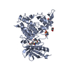
 PDBj
PDBj
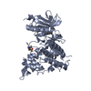
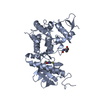
 Assembly
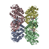

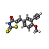
 Mass: 275.346 Da / Num. of mol.: 4 / Source method: obtained synthetically / Formula: C13H9NO2S2 / Feature type: SUBJECT OF INVESTIGATION
 Mass: 18.015 Da / Num. of mol.: 159 / Source method: isolated from a natural source / Formula: H2O
 Sample preparation
 Processing