 Movie
Movie Controller

[English] 日本語
 Yorodumi
- PDB-7ebh: Crystal structure of human pyruvate dehydrogenase kinase 2 in com... -
+ Open data

- Basic information
| Entry | Database: PDB / ID: 7ebh | ||||||
|---|---|---|---|---|---|---|---|
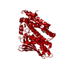
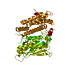
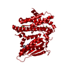
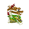
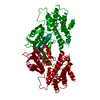
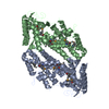
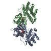
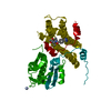
| Title | Crystal structure of human pyruvate dehydrogenase kinase 2 in complex with compound 13 | ||||||
 Components Components | [Pyruvate dehydrogenase (acetyl-transferring)] kinase isozyme 2, mitochondrial | ||||||
 Keywords Keywords | TRANSFERASE / PDHK / KINASE INHIBITORS / FRAGMENT SCREENING / PDK1 / PDK2 / PDK3 / PDK4 | ||||||
| Function / homology |  Function and homology information Function and homology information[pyruvate dehydrogenase (acetyl-transferring)] kinase / pyruvate dehydrogenase (acetyl-transferring) kinase activity / regulation of pyruvate decarboxylation to acetyl-CoA / Regulation of pyruvate dehydrogenase (PDH) complex / pyruvate dehydrogenase complex / regulation of pH / regulation of ketone metabolic process / cellular response to nutrient / regulation of gluconeogenesis / Signaling by Retinoic Acid ...[pyruvate dehydrogenase (acetyl-transferring)] kinase / pyruvate dehydrogenase (acetyl-transferring) kinase activity / regulation of pyruvate decarboxylation to acetyl-CoA / Regulation of pyruvate dehydrogenase (PDH) complex / pyruvate dehydrogenase complex / regulation of pH / regulation of ketone metabolic process / cellular response to nutrient / regulation of gluconeogenesis / Signaling by Retinoic Acid / intrinsic apoptotic signaling pathway by p53 class mediator / regulation of glucose metabolic process / cellular response to reactive oxygen species / glucose metabolic process / glucose homeostasis / insulin receptor signaling pathway / protein kinase activity / mitochondrial matrix / protein homodimerization activity / mitochondrion / ATP binding Similarity search - Function | ||||||
| Biological species |  Homo sapiens (human) Homo sapiens (human) | ||||||
| Method |  X-RAY DIFFRACTION / SYNCHROTRON / MOLECULAR REPLACEMENT / Resolution: 1.96 Å X-RAY DIFFRACTION / SYNCHROTRON / MOLECULAR REPLACEMENT / Resolution: 1.96 Å | ||||||
 Authors Authors | Orita, T. / Doi, S. / Iwanaga, T. / Adachi, T. | ||||||
 Citation Citation | Journal: Bioorg.Med.Chem. / Year: 2021 Title: Fragment-based lead discovery to identify novel inhibitors that target the ATP binding site of pyruvate dehydrogenase kinases. Authors: Akaki, T. / Bessho, Y. / Ito, T. / Fujioka, S. / Ubukata, M. / Mori, G. / Yamanaka, K. / Orita, T. / Doi, S. / Iwanaga, T. / Ikegashira, K. / Hantani, Y. / Nakanishi, I. / Adachi, T. | ||||||
| History |
|
- Structure visualization
| Structure viewer | Molecule: MolmilJmol/JSmol |
|---|
- Downloads & links
-Download
| PDBx/mmCIF format | 7ebh.cif.gz | 178.1 KB | Display | PDBx/mmCIF format |
|---|---|---|---|---|
| PDB format | pdb7ebh.ent.gz | 125.2 KB | Display | PDB format |
| PDBx/mmJSON format | 7ebh.json.gz | Tree view | PDBx/mmJSON format | |
| Others |  Other downloads Other downloads |
-Validation report
| Arichive directory | https://data.pdbj.org/pub/pdb/validation_reports/eb/7ebhftp://data.pdbj.org/pub/pdb/validation_reports/eb/7ebh | HTTPS FTP |
|---|
-Related structure data
| Related structure data |  7ea0C  7easC  7eatC  7ebbC 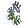 7ebgC  2btzS C: citing same article ( S: Starting model for refinement |
|---|---|
| Similar structure data |


-Links
 PDBj
PDBj


- Assembly
| Deposited unit | 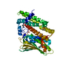
| ||||||||||||
|---|---|---|---|---|---|---|---|---|---|---|---|---|---|
| 1 | 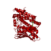
| ||||||||||||
| Unit cell |
|
-Components
-Protein , 1 types, 1 molecules A
| #1: Protein | [ Mass: 44647.730 Da / Num. of mol.: 1 Source method: isolated from a genetically manipulated source Source: (gene. exp.) Homo sapiens (human) / Gene: PDK2, PDHK2 / Production host:  References: UniProt: Q15119, [pyruvate dehydrogenase (acetyl-transferring)] kinase |
|---|
-Non-polymers , 5 types, 94 molecules 


| #2: Chemical | ChemComp-CL /  Mass: 35.453 Da / Num. of mol.: 1 / Source method: obtained synthetically / Formula: Cl Mass: 35.453 Da / Num. of mol.: 1 / Source method: obtained synthetically / Formula: Cl | ||||||
|---|---|---|---|---|---|---|---|
| #3: Chemical |  Mass: 59.044 Da / Num. of mol.: 2 / Source method: obtained synthetically / Formula: C2H3O2 Mass: 59.044 Da / Num. of mol.: 2 / Source method: obtained synthetically / Formula: C2H3O2#4: Chemical |  Mass: 78.133 Da / Num. of mol.: 2 / Source method: obtained synthetically / Formula: C2H6OS / Comment: DMSO, precipitant*YM Mass: 78.133 Da / Num. of mol.: 2 / Source method: obtained synthetically / Formula: C2H6OS / Comment: DMSO, precipitant*YM#5: Chemical | ChemComp-J0F / |  Mass: 254.126 Da / Num. of mol.: 1 / Source method: obtained synthetically / Formula: C10H12BrN3 / Feature type: SUBJECT OF INVESTIGATION Mass: 254.126 Da / Num. of mol.: 1 / Source method: obtained synthetically / Formula: C10H12BrN3 / Feature type: SUBJECT OF INVESTIGATION#6: Water | ChemComp-HOH / | Mass: 18.015 Da / Num. of mol.: 88 / Source method: isolated from a natural source / Formula: H2O |
-Details
| Has ligand of interest | Y |
|---|
-Experimental details
-Experiment
| Experiment | Method: X-RAY DIFFRACTION / Number of used crystals: 1 |
|---|
- Sample preparation
| Crystal | Density Matthews: 3.25 Å3/Da / Density % sol: 62.19 % |
|---|---|
| Crystal grow | Temperature: 277.15 K / Method: vapor diffusion, hanging drop / pH: 5.5 Details: 50 mM Na acetate pH 5.5, 100 mM magnesium chloride and 8% (v/v) isopropanol |
-Data collection
| Diffraction | Mean temperature: 100 K / Serial crystal experiment: N |
|---|---|
| Diffraction source | Source: SYNCHROTRON / Site: SPring-8  / Beamline: BL41XU / Wavelength: 1 Å / Beamline: BL41XU / Wavelength: 1 Å |
| Detector | Type: DECTRIS PILATUS3 S 6M / Detector: PIXEL / Date: Jul 23, 2015 |
| Radiation | Protocol: SINGLE WAVELENGTH / Monochromatic (M) / Laue (L): M / Scattering type: x-ray |
| Radiation wavelength | Wavelength: 1 Å / Relative weight: 1 |
| Reflection | Resolution: 1.96→94.7 Å / Num. obs: 40300 / % possible obs: 98.7 % / Redundancy: 10.1 % / Biso Wilson estimate: 41.69 Å2 / CC1/2: 0.999 / Rmerge(I) obs: 0.056 / Rpim(I) all: 0.018 / Rrim(I) all: 0.059 / Net I/σ(I): 19.5 |
| Reflection shell | Resolution: 1.96→2.02 Å / Redundancy: 10 % / Rmerge(I) obs: 1.015 / Mean I/σ(I) obs: 2.6 / Num. unique obs: 2943 / CC1/2: 0.802 / Rpim(I) all: 0.333 / Rrim(I) all: 1.069 / % possible all: 97.7 |
- Processing
| Software |
| ||||||||||||||||||||||||||||||||||||||||||||||||||||||||||||||||||||||||||||||||||||||||||||||||||||||||||||||||
|---|---|---|---|---|---|---|---|---|---|---|---|---|---|---|---|---|---|---|---|---|---|---|---|---|---|---|---|---|---|---|---|---|---|---|---|---|---|---|---|---|---|---|---|---|---|---|---|---|---|---|---|---|---|---|---|---|---|---|---|---|---|---|---|---|---|---|---|---|---|---|---|---|---|---|---|---|---|---|---|---|---|---|---|---|---|---|---|---|---|---|---|---|---|---|---|---|---|---|---|---|---|---|---|---|---|---|---|---|---|---|---|---|---|
| Refinement | Method to determine structure: MOLECULAR REPLACEMENT Starting model: 2btz Resolution: 1.96→94.7 Å / SU ML: 0.2166 / Cross valid method: FREE R-VALUE / σ(F): 1.34 / Phase error: 22.5776 Stereochemistry target values: GeoStd + Monomer Library + CDL v1.2
| ||||||||||||||||||||||||||||||||||||||||||||||||||||||||||||||||||||||||||||||||||||||||||||||||||||||||||||||||
| Solvent computation | Shrinkage radii: 0.9 Å / VDW probe radii: 1.11 Å / Solvent model: FLAT BULK SOLVENT MODEL | ||||||||||||||||||||||||||||||||||||||||||||||||||||||||||||||||||||||||||||||||||||||||||||||||||||||||||||||||
| Displacement parameters | Biso mean: 53.63 Å2 | ||||||||||||||||||||||||||||||||||||||||||||||||||||||||||||||||||||||||||||||||||||||||||||||||||||||||||||||||
| Refinement step | Cycle: LAST / Resolution: 1.96→94.7 Å
| ||||||||||||||||||||||||||||||||||||||||||||||||||||||||||||||||||||||||||||||||||||||||||||||||||||||||||||||||
| Refine LS restraints |
| ||||||||||||||||||||||||||||||||||||||||||||||||||||||||||||||||||||||||||||||||||||||||||||||||||||||||||||||||
| LS refinement shell |
| ||||||||||||||||||||||||||||||||||||||||||||||||||||||||||||||||||||||||||||||||||||||||||||||||||||||||||||||||
| Refinement TLS params. | Method: refined / Origin x: 46.7495288993 Å / Origin y: 45.6732922148 Å / Origin z: 72.4035994514 Å
| ||||||||||||||||||||||||||||||||||||||||||||||||||||||||||||||||||||||||||||||||||||||||||||||||||||||||||||||||
| Refinement TLS group | Selection details: all |
