 Movie
Movie Controller

[English] 日本語
 Yorodumi
- PDB-7e1p: Crystal structure of Sulfurisphaera tokodaii O6-methylguanine met... -
+ Open data

- Basic information
| Entry | Database: PDB / ID: 7e1p | ||||||
|---|---|---|---|---|---|---|---|
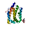
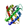
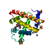
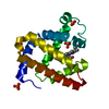
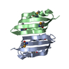
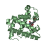
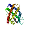
| Title | Crystal structure of Sulfurisphaera tokodaii O6-methylguanine methyltransferase C120S variant in complex with O6-methyldeoxyguanosine | ||||||
 Components Components | Methylated-DNA--protein-cysteine methyltransferase | ||||||
 Keywords Keywords | TRANSFERASE / methyltransferase | ||||||
| Function / homology |  Function and homology information Function and homology informationmethylated-DNA-[protein]-cysteine S-methyltransferase / methylated-DNA-[protein]-cysteine S-methyltransferase activity / DNA alkylation repair / methylation / cytoplasm Similarity search - Function | ||||||
| Biological species |   Sulfurisphaera tokodaii (archaea) Sulfurisphaera tokodaii (archaea) | ||||||
| Method |  X-RAY DIFFRACTION / SYNCHROTRON / MOLECULAR REPLACEMENT / Resolution: 1.63 Å X-RAY DIFFRACTION / SYNCHROTRON / MOLECULAR REPLACEMENT / Resolution: 1.63 Å | ||||||
 Authors Authors | Kikuchi, M. / Yamauchi, T. / Iizuka, Y. / Tsunoda, M. | ||||||
 Citation Citation | Journal: Acta Crystallogr.,Sect.F / Year: 2021 Title: Roles of the hydroxy group of tyrosine in crystal structures of Sulfurisphaera tokodaii O6-methylguanine-DNA methyltransferase. Authors: Kikuchi, M. / Yamauchi, T. / Iizuka, Y. / Tsunoda, M. | ||||||
| History |
|
- Structure visualization
| Structure viewer | Molecule: MolmilJmol/JSmol |
|---|
- Downloads & links
-Download
| PDBx/mmCIF format | 7e1p.cif.gz | 52.4 KB | Display | PDBx/mmCIF format |
|---|---|---|---|---|
| PDB format | pdb7e1p.ent.gz | 34.1 KB | Display | PDB format |
| PDBx/mmJSON format | 7e1p.json.gz | Tree view | PDBx/mmJSON format | |
| Others |  Other downloads Other downloads |
-Validation report
| Arichive directory | https://data.pdbj.org/pub/pdb/validation_reports/e1/7e1pftp://data.pdbj.org/pub/pdb/validation_reports/e1/7e1p | HTTPS FTP |
|---|
-Related structure data
| Related structure data |  7csmC  7d4vC  7dknC  7dqqC  7dqrC 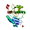 7dqtC  1wrjS S: Starting model for refinement C: citing same article ( |
|---|---|
| Similar structure data |


-Links
 PDBj
PDBj- Assembly
| Deposited unit | 
| ||||||||
|---|---|---|---|---|---|---|---|---|---|
| 1 |
| ||||||||
| Unit cell |
|
-Components
| #1: Protein | Mass: 17960.887 Da / Num. of mol.: 1 / Mutation: C120S Source method: isolated from a genetically manipulated source Source: (gene. exp.) Sulfurisphaera tokodaii (strain DSM 16993 / JCM 10545 / NBRC 100140 / 7) (archaea)Strain: DSM 16993 / JCM 10545 / NBRC 100140 / 7 / Gene: ogt, STK_09670 / Production host:  References: UniProt: Q973C7, methylated-DNA-[protein]-cysteine S-methyltransferase |
|---|---|
| #2: Chemical | ChemComp-J03 / (  Mass: 281.268 Da / Num. of mol.: 1 / Source method: obtained synthetically / Formula: C11H15N5O4 / Feature type: SUBJECT OF INVESTIGATION Mass: 281.268 Da / Num. of mol.: 1 / Source method: obtained synthetically / Formula: C11H15N5O4 / Feature type: SUBJECT OF INVESTIGATION |
| #3: Chemical | ChemComp-SO4 /   Mass: 96.063 Da / Num. of mol.: 1 / Source method: obtained synthetically / Formula: SO4 Mass: 96.063 Da / Num. of mol.: 1 / Source method: obtained synthetically / Formula: SO4 |
| #4: Water | ChemComp-HOH /  Mass: 18.015 Da / Num. of mol.: 145 / Source method: isolated from a natural source / Formula: H2O Mass: 18.015 Da / Num. of mol.: 145 / Source method: isolated from a natural source / Formula: H2O |
| Has ligand of interest | Y |
| Has protein modification | Y |
-Experimental details
-Experiment
| Experiment | Method: X-RAY DIFFRACTION / Number of used crystals: 1 |
|---|
- Sample preparation
| Crystal | Density Matthews: 2.19 Å3/Da / Density % sol: 43.71 % |
|---|---|
| Crystal grow | Temperature: 293 K / Method: vapor diffusion, hanging drop Details: 0.2 M Ammonium sulfate, 0.1 M Tris pH 8.5, 25% w/v Polyethylene glycol 3,350 |
-Data collection
| Diffraction | Mean temperature: 100 K / Serial crystal experiment: N | ||||||||||||||||||||||||||||||
|---|---|---|---|---|---|---|---|---|---|---|---|---|---|---|---|---|---|---|---|---|---|---|---|---|---|---|---|---|---|---|---|
| Diffraction source | Source: SYNCHROTRON / Site: Photon Factory  / Beamline: BL-5A / Wavelength: 1 Å / Beamline: BL-5A / Wavelength: 1 Å | ||||||||||||||||||||||||||||||
| Detector | Type: DECTRIS PILATUS3 S 6M / Detector: PIXEL / Date: May 19, 2019 | ||||||||||||||||||||||||||||||
| Radiation | Protocol: SINGLE WAVELENGTH / Monochromatic (M) / Laue (L): M / Scattering type: x-ray | ||||||||||||||||||||||||||||||
| Radiation wavelength | Wavelength: 1 Å / Relative weight: 1 | ||||||||||||||||||||||||||||||
| Reflection | Resolution: 1.63→48.33 Å / Num. obs: 20281 / % possible obs: 100 % / Redundancy: 6.4 % / CC1/2: 1 / Rmerge(I) obs: 0.025 / Rpim(I) all: 0.011 / Rrim(I) all: 0.027 / Net I/σ(I): 40 / Num. measured all: 129783 | ||||||||||||||||||||||||||||||
| Reflection shell | Diffraction-ID: 1
|
- Processing
| Software |
| ||||||||||||||||||||||||||||||||||||||||||||||||||||||||||||||||||||||||||||||||||||||||||||||||||||||||||||||||||||||||||||||||||||||||||||||||||||||||||||||||
|---|---|---|---|---|---|---|---|---|---|---|---|---|---|---|---|---|---|---|---|---|---|---|---|---|---|---|---|---|---|---|---|---|---|---|---|---|---|---|---|---|---|---|---|---|---|---|---|---|---|---|---|---|---|---|---|---|---|---|---|---|---|---|---|---|---|---|---|---|---|---|---|---|---|---|---|---|---|---|---|---|---|---|---|---|---|---|---|---|---|---|---|---|---|---|---|---|---|---|---|---|---|---|---|---|---|---|---|---|---|---|---|---|---|---|---|---|---|---|---|---|---|---|---|---|---|---|---|---|---|---|---|---|---|---|---|---|---|---|---|---|---|---|---|---|---|---|---|---|---|---|---|---|---|---|---|---|---|---|---|---|---|
| Refinement | Method to determine structure: MOLECULAR REPLACEMENT Starting model: 1wrj Resolution: 1.63→30.859 Å / Cor.coef. Fo:Fc: 0.959 / Cor.coef. Fo:Fc free: 0.937 / SU B: 1.676 / SU ML: 0.059 / Cross valid method: FREE R-VALUE / ESU R: 0.097 / ESU R Free: 0.1 Details: Hydrogens have been added in their riding positions
| ||||||||||||||||||||||||||||||||||||||||||||||||||||||||||||||||||||||||||||||||||||||||||||||||||||||||||||||||||||||||||||||||||||||||||||||||||||||||||||||||
| Solvent computation | Ion probe radii: 0.8 Å / Shrinkage radii: 0.8 Å / VDW probe radii: 1.2 Å / Solvent model: MASK BULK SOLVENT | ||||||||||||||||||||||||||||||||||||||||||||||||||||||||||||||||||||||||||||||||||||||||||||||||||||||||||||||||||||||||||||||||||||||||||||||||||||||||||||||||
| Displacement parameters | Biso mean: 19.259 Å2
| ||||||||||||||||||||||||||||||||||||||||||||||||||||||||||||||||||||||||||||||||||||||||||||||||||||||||||||||||||||||||||||||||||||||||||||||||||||||||||||||||
| Refinement step | Cycle: LAST / Resolution: 1.63→30.859 Å
| ||||||||||||||||||||||||||||||||||||||||||||||||||||||||||||||||||||||||||||||||||||||||||||||||||||||||||||||||||||||||||||||||||||||||||||||||||||||||||||||||
| Refine LS restraints |
| ||||||||||||||||||||||||||||||||||||||||||||||||||||||||||||||||||||||||||||||||||||||||||||||||||||||||||||||||||||||||||||||||||||||||||||||||||||||||||||||||
| LS refinement shell |
|
