 Movie
Movie Controller

[English] 日本語
 Yorodumi
- PDB-7doy: The structure of the Arabidopsis thaliana guanosine deaminase in ... -
+ Open data

- Basic information
| Entry | Database: PDB / ID: 7doy | ||||||
|---|---|---|---|---|---|---|---|
| Title | The structure of the Arabidopsis thaliana guanosine deaminase in complex with 6-O-methylguanosine | ||||||
 Components Components | Guanosine deaminase | ||||||
 Keywords Keywords | PLANT PROTEIN / deamination / GSDA / purine metabolism | ||||||
| Function / homology |  Function and homology information Function and homology informationguanosine deaminase / guanosine deaminase activity / purine nucleoside catabolic process / zinc ion binding / nucleus / cytoplasm Similarity search - Function | ||||||
| Biological species |  | ||||||
| Method |  X-RAY DIFFRACTION / SYNCHROTRON / MOLECULAR REPLACEMENT / Resolution: 2.17 Å X-RAY DIFFRACTION / SYNCHROTRON / MOLECULAR REPLACEMENT / Resolution: 2.17 Å | ||||||
 Authors Authors | Xie, W. / Jia, Q. / Zeng, H. | ||||||
| Funding support |  China, 1items China, 1items
| ||||||
 Citation Citation | Journal: To Be Published Title: Asymmetric Catalysis of Arabidopsis thaliana Guanosine Deaminase Revealed by Crystal Structures Authors: Xie, W. / Jia, Q. / Zeng, H. | ||||||
| History |
|
- Structure visualization
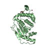
| Structure viewer | Molecule: MolmilJmol/JSmol |
|---|
- Downloads & links
-Download
| PDBx/mmCIF format | 7doy.cif.gz | 92.6 KB | Display | PDBx/mmCIF format |
|---|---|---|---|---|
| PDB format | pdb7doy.ent.gz | 56.6 KB | Display | PDB format |
| PDBx/mmJSON format | 7doy.json.gz | Tree view | PDBx/mmJSON format | |
| Others |  Other downloads Other downloads |
-Validation report
| Summary document | 7doy_validation.pdf.gz | 2.3 MB | Display | wwPDB validaton report |
|---|---|---|---|---|
| Full document | 7doy_full_validation.pdf.gz | 2.3 MB | Display | |
| Data in XML | 7doy_validation.xml.gz | 16 KB | Display | |
| Data in CIF | 7doy_validation.cif.gz | 21.5 KB | Display | |
| Arichive directory | https://data.pdbj.org/pub/pdb/validation_reports/do/7doyftp://data.pdbj.org/pub/pdb/validation_reports/do/7doy | HTTPS FTP |
-Related structure data
| Related structure data |  7dlcC  7dowC  7doxC  7dpkC  7dbfS S: Starting model for refinement C: citing same article ( |
|---|---|
| Similar structure data |


-Links
 PDBj
PDBj- Assembly
| Deposited unit | 
| ||||||||||||
|---|---|---|---|---|---|---|---|---|---|---|---|---|---|
| 1 |
| ||||||||||||
| Unit cell |
|
-Components
| #1: Protein | Mass: 17523.016 Da / Num. of mol.: 2 Source method: isolated from a genetically manipulated source Source: (gene. exp.)  #2: Chemical |   Mass: 297.267 Da / Num. of mol.: 2 / Source method: obtained synthetically / Formula: C11H15N5O5 / Feature type: SUBJECT OF INVESTIGATION Mass: 297.267 Da / Num. of mol.: 2 / Source method: obtained synthetically / Formula: C11H15N5O5 / Feature type: SUBJECT OF INVESTIGATION#3: Chemical |   Mass: 65.409 Da / Num. of mol.: 2 / Source method: obtained synthetically / Formula: Zn / Feature type: SUBJECT OF INVESTIGATION Mass: 65.409 Da / Num. of mol.: 2 / Source method: obtained synthetically / Formula: Zn / Feature type: SUBJECT OF INVESTIGATION#4: Water | ChemComp-HOH / |  Mass: 18.015 Da / Num. of mol.: 152 / Source method: isolated from a natural source / Formula: H2O Mass: 18.015 Da / Num. of mol.: 152 / Source method: isolated from a natural source / Formula: H2OHas ligand of interest | Y | |
|---|
-Experimental details
-Experiment
| Experiment | Method: X-RAY DIFFRACTION / Number of used crystals: 1 |
|---|
- Sample preparation
| Crystal | Density Matthews: 2.32 Å3/Da / Density % sol: 47.04 % |
|---|---|
| Crystal grow | Temperature: 277 K / Method: vapor diffusion, sitting drop / pH: 7.5 / Details: 1.2 M Na-citrate and 0.1 M HEPES (pH 7.5) |
-Data collection
| Diffraction | Mean temperature: 100 K / Serial crystal experiment: N |
|---|---|
| Diffraction source | Source: SYNCHROTRON / Site: SSRF / Beamline: BL19U1 / Wavelength: 0.979 Å |
| Detector | Type: DECTRIS PILATUS3 S 6M / Detector: PIXEL / Date: Apr 17, 2020 |
| Radiation | Protocol: SINGLE WAVELENGTH / Monochromatic (M) / Laue (L): M / Scattering type: x-ray |
| Radiation wavelength | Wavelength: 0.979 Å / Relative weight: 1 |
| Reflection | Resolution: 2.17→50 Å / Num. obs: 17386 / % possible obs: 100 % / Redundancy: 19.8 % / Biso Wilson estimate: 29.92 Å2 / CC1/2: 0.992 / Rmerge(I) obs: 0.177 / Rrim(I) all: 0.18 / Net I/σ(I): 25.6 |
| Reflection shell | Resolution: 2.17→2.25 Å / Redundancy: 17.7 % / Rmerge(I) obs: 0.501 / Mean I/σ(I) obs: 6.333 / Num. unique obs: 1711 / CC1/2: 0.952 / % possible all: 100 |
- Processing
| Software |
| |||||||||||||||||||||||||||||||||||||||||||||||||
|---|---|---|---|---|---|---|---|---|---|---|---|---|---|---|---|---|---|---|---|---|---|---|---|---|---|---|---|---|---|---|---|---|---|---|---|---|---|---|---|---|---|---|---|---|---|---|---|---|---|---|
| Refinement | Method to determine structure: MOLECULAR REPLACEMENT Starting model: 7DBF Resolution: 2.17→28.61 Å / SU ML: 0.1931 / Cross valid method: FREE R-VALUE / σ(F): 1.38 / Phase error: 21.8036 / Stereochemistry target values: CDL v1.2
| |||||||||||||||||||||||||||||||||||||||||||||||||
| Solvent computation | Shrinkage radii: 0.9 Å / VDW probe radii: 1.11 Å / Solvent model: FLAT BULK SOLVENT MODEL | |||||||||||||||||||||||||||||||||||||||||||||||||
| Displacement parameters | Biso mean: 35.7 Å2 | |||||||||||||||||||||||||||||||||||||||||||||||||
| Refinement step | Cycle: LAST / Resolution: 2.17→28.61 Å
| |||||||||||||||||||||||||||||||||||||||||||||||||
| Refine LS restraints |
| |||||||||||||||||||||||||||||||||||||||||||||||||
| LS refinement shell |
|
