 Movie
Movie Controller

[English] 日本語
 Yorodumi
- PDB-7d2k: Crystal structure of rat TRPV6 in complex with (4- phenylcyclohex... -
+ Open data

- Basic information
| Entry | Database: PDB / ID: 7d2k | ||||||
|---|---|---|---|---|---|---|---|
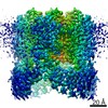
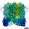
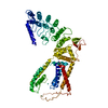
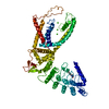
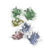
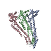
| Title | Crystal structure of rat TRPV6 in complex with (4- phenylcyclohexyl)piperazine inhibitor Br-cis-22a | ||||||
 Components Components | Transient receptor potential cation channel subfamily V member 6 | ||||||
 Keywords Keywords | TRANSPORT PROTEIN/INHIBITOR / Ion channels / TRP channels / Membrane proteins / TRANSPORT PROTEIN-INHIBITOR complex | ||||||
| Function / homology |  Function and homology information Function and homology informationparathyroid hormone secretion / TRP channels / calcium ion import / calcium-activated cation channel activity / calcium ion import across plasma membrane / calcium ion homeostasis / calcium channel complex / response to calcium ion / calcium ion transmembrane transport / calcium channel activity ...parathyroid hormone secretion / TRP channels / calcium ion import / calcium-activated cation channel activity / calcium ion import across plasma membrane / calcium ion homeostasis / calcium channel complex / response to calcium ion / calcium ion transmembrane transport / calcium channel activity / calcium ion transport / protein homotetramerization / calmodulin binding / apical plasma membrane / metal ion binding / identical protein binding / plasma membrane Similarity search - Function | ||||||
| Biological species |  | ||||||
| Method |  X-RAY DIFFRACTION / SYNCHROTRON / MOLECULAR REPLACEMENT / Resolution: 3.698 Å X-RAY DIFFRACTION / SYNCHROTRON / MOLECULAR REPLACEMENT / Resolution: 3.698 Å | ||||||
 Authors Authors | Singh, A.K. / Neuberger, A. / Nadezhdin, K.D. / Sobolevsky, A.I. | ||||||
| Funding support |  United States, 1items United States, 1items
| ||||||
 Citation Citation | Journal: Sci Adv / Year: 2020 Title: Inactivation-mimicking block of the epithelial calcium channel TRPV6. Authors: Rajesh Bhardwaj / Sonja Lindinger / Arthur Neuberger / Kirill D Nadezhdin / Appu K Singh / Micael R Cunha / Isabella Derler / Gergely Gyimesi / Jean-Louis Reymond / Matthias A Hediger / ...Authors: Rajesh Bhardwaj / Sonja Lindinger / Arthur Neuberger / Kirill D Nadezhdin / Appu K Singh / Micael R Cunha / Isabella Derler / Gergely Gyimesi / Jean-Louis Reymond / Matthias A Hediger / Christoph Romanin / Alexander I Sobolevsky /    Abstract: Epithelial calcium channel TRPV6 plays vital roles in calcium homeostasis, and its dysregulation is implicated in multifactorial diseases, including cancers. Here, we study the molecular mechanism of ...Epithelial calcium channel TRPV6 plays vital roles in calcium homeostasis, and its dysregulation is implicated in multifactorial diseases, including cancers. Here, we study the molecular mechanism of selective nanomolar-affinity TRPV6 inhibition by (4-phenylcyclohexyl)piperazine derivatives (PCHPDs). We use x-ray crystallography and cryo-electron microscopy to solve the inhibitor-bound structures of TRPV6 and identify two types of inhibitor binding sites in the transmembrane region: (i) modulatory sites between the S1-S4 and pore domains normally occupied by lipids and (ii) the main site in the ion channel pore. Our structural data combined with mutagenesis, functional and computational approaches suggest that PCHPDs plug the open pore of TRPV6 and convert the channel into a nonconducting state, mimicking the action of calmodulin, which causes inactivation of TRPV6 channels under physiological conditions. This mechanism of inhibition explains the high selectivity and potency of PCHPDs and opens up unexplored avenues for the design of future-generation biomimetic drugs. | ||||||
| History |
|
- Structure visualization
| Structure viewer | Molecule: MolmilJmol/JSmol |
|---|
- Downloads & links
-Download
| PDBx/mmCIF format | 7d2k.cif.gz | 135 KB | Display | PDBx/mmCIF format |
|---|---|---|---|---|
| PDB format | pdb7d2k.ent.gz | 102.5 KB | Display | PDB format |
| PDBx/mmJSON format | 7d2k.json.gz | Tree view | PDBx/mmJSON format | |
| Others |  Other downloads Other downloads |
-Validation report
| Arichive directory | https://data.pdbj.org/pub/pdb/validation_reports/d2/7d2kftp://data.pdbj.org/pub/pdb/validation_reports/d2/7d2k | HTTPS FTP |
|---|
-Related structure data
| Related structure data |  7k4aC  7k4bC  7k4cC  7k4dC 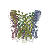 7k4eC 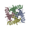 7k4fC  5wo7S S: Starting model for refinement C: citing same article ( |
|---|---|
| Similar structure data |


-Links
 PDBj
PDBj
- Assembly
| Deposited unit | 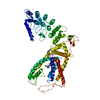
| ||||||||||||||||||
|---|---|---|---|---|---|---|---|---|---|---|---|---|---|---|---|---|---|---|---|
| 1 |
| ||||||||||||||||||
| Unit cell |
| ||||||||||||||||||
| Components on special symmetry positions |
|
-Components
| #1: Protein | Mass: 77133.492 Da / Num. of mol.: 1 / Fragment: UNP residues 41-709 / Mutation: I62Y, L132N, M96Q Source method: isolated from a genetically manipulated source Source: (gene. exp.)  Homo sapiens (human) / References: UniProt: Q9R186 Homo sapiens (human) / References: UniProt: Q9R186 | ||
|---|---|---|---|
| #2: Chemical | ChemComp-CA /   Mass: 40.078 Da / Num. of mol.: 1 / Source method: obtained synthetically / Formula: Ca Mass: 40.078 Da / Num. of mol.: 1 / Source method: obtained synthetically / Formula: Ca | ||
| #3: Chemical |   Mass: 415.390 Da / Num. of mol.: 2 / Source method: obtained synthetically / Formula: C22H29BrN3 / Feature type: SUBJECT OF INVESTIGATION Mass: 415.390 Da / Num. of mol.: 2 / Source method: obtained synthetically / Formula: C22H29BrN3 / Feature type: SUBJECT OF INVESTIGATIONHas ligand of interest | Y | |
-Experimental details
-Experiment
| Experiment | Method: X-RAY DIFFRACTION / Number of used crystals: 1 |
|---|
- Sample preparation
| Crystal | Density Matthews: 3.94 Å3/Da / Density % sol: 68.78 % |
|---|---|
| Crystal grow | Temperature: 293 K / Method: vapor diffusion, hanging drop / pH: 8 / Details: 150 mM nacl, 100 mM TRIS, 22% PEG 350 / PH range: 8.0-8.5 |
-Data collection
| Diffraction | Mean temperature: 100 K / Serial crystal experiment: N |
|---|---|
| Diffraction source | Source: SYNCHROTRON / Site: APS / Beamline: 24-ID-C / Wavelength: 0.92 Å |
| Detector | Type: DECTRIS PILATUS 6M-F / Detector: PIXEL / Date: Mar 12, 2019 |
| Radiation | Monochromator: M / Protocol: SINGLE WAVELENGTH / Monochromatic (M) / Laue (L): M / Scattering type: x-ray |
| Radiation wavelength | Wavelength: 0.92 Å / Relative weight: 1 |
| Reflection | Resolution: 3.6→47.129 Å / Num. obs: 14416 / % possible obs: 99.12 % / Redundancy: 8.13 % / CC1/2: 0.99 / Net I/σ(I): 1.45 |
| Reflection shell | Resolution: 3.6→3.9 Å / Num. unique obs: 2587 / CC1/2: 0.61 / % possible all: 96.2 |
- Processing
| Software |
| ||||||||||||||||||||||||||||||||||||||||||
|---|---|---|---|---|---|---|---|---|---|---|---|---|---|---|---|---|---|---|---|---|---|---|---|---|---|---|---|---|---|---|---|---|---|---|---|---|---|---|---|---|---|---|---|
| Refinement | Method to determine structure: MOLECULAR REPLACEMENT Starting model: 5WO7 Resolution: 3.698→47.12 Å / SU ML: 0.76 / Cross valid method: FREE R-VALUE / σ(F): 1.36 / Phase error: 48.89 / Stereochemistry target values: ML
| ||||||||||||||||||||||||||||||||||||||||||
| Solvent computation | Shrinkage radii: 0.9 Å / VDW probe radii: 1.11 Å / Solvent model: FLAT BULK SOLVENT MODEL | ||||||||||||||||||||||||||||||||||||||||||
| Refinement step | Cycle: LAST / Resolution: 3.698→47.12 Å
| ||||||||||||||||||||||||||||||||||||||||||
| Refine LS restraints |
| ||||||||||||||||||||||||||||||||||||||||||
| LS refinement shell |
|
