 Movie
Movie Controller

[English] 日本語
 Yorodumi
- PDB-7bxr: 2-amino-3-ketobutyrate CoA ligase from Cupriavidus necator 3-Hydr... -
+ Open data

- Basic information
| Entry | Database: PDB / ID: 7bxr | |||||||||
|---|---|---|---|---|---|---|---|---|---|---|
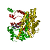
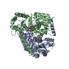
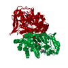
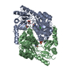
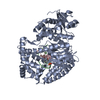
| Title | 2-amino-3-ketobutyrate CoA ligase from Cupriavidus necator 3-Hydroxynorvaline binding form | |||||||||
 Components Components | 2-amino-3-ketobutyrate coenzyme A ligase | |||||||||
 Keywords Keywords | LIGASE / TRANSFERASE / 2-amino-3-ketobutyrate CoA ligase / Cupriavidus necator | |||||||||
| Function / homology |  Function and homology information Function and homology informationglycine C-acetyltransferase / glycine C-acetyltransferase activity / L-threonine catabolic process to glycine / biosynthetic process / ligase activity / pyridoxal phosphate binding / cytosol Similarity search - Function | |||||||||
| Biological species |  Cupriavidus necator (bacteria) Cupriavidus necator (bacteria) | |||||||||
| Method |  X-RAY DIFFRACTION / SYNCHROTRON / MOLECULAR REPLACEMENT / Resolution: 2.55 Å X-RAY DIFFRACTION / SYNCHROTRON / MOLECULAR REPLACEMENT / Resolution: 2.55 Å | |||||||||
 Authors Authors | Motoyama, T. / Nakano, S. / Hasebe, F. / Miyoshi, N. / Ito, S. | |||||||||
| Funding support |  Japan, 2items Japan, 2items
| |||||||||
 Citation Citation | Journal: Commun Chem / Year: 2021 Title: Chemoenzymatic synthesis of 3-ethyl-2,5-dimethylpyrazine by L-threonine 3-dehydrogenase and 2-amino-3-ketobutyrate CoA ligase/L-threonine aldolase Authors: Motoyama, T. / Nakano, S. / Hasebe, F. / Miyata, R. / Kumazawa, S. / Miyoshi, N. / Ito, S. | |||||||||
| History |
|
- Structure visualization
| Structure viewer | Molecule: MolmilJmol/JSmol |
|---|
- Downloads & links
-Download
| PDBx/mmCIF format | 7bxr.cif.gz | 205.6 KB | Display | PDBx/mmCIF format |
|---|---|---|---|---|
| PDB format | pdb7bxr.ent.gz | 130.9 KB | Display | PDB format |
| PDBx/mmJSON format | 7bxr.json.gz | Tree view | PDBx/mmJSON format | |
| Others |  Other downloads Other downloads |
-Validation report
| Summary document | 7bxr_validation.pdf.gz | 1.1 MB | Display | wwPDB validaton report |
|---|---|---|---|---|
| Full document | 7bxr_full_validation.pdf.gz | 1.1 MB | Display | |
| Data in XML | 7bxr_validation.xml.gz | 33.3 KB | Display | |
| Data in CIF | 7bxr_validation.cif.gz | 46.3 KB | Display | |
| Arichive directory | https://data.pdbj.org/pub/pdb/validation_reports/bx/7bxrftp://data.pdbj.org/pub/pdb/validation_reports/bx/7bxr | HTTPS FTP |
-Related structure data
| Related structure data |  7bxpC  7bxqC  7bxsC  1fc4S S: Starting model for refinement C: citing same article ( |
|---|---|
| Similar structure data |

-Links
 PDBj
PDBj- Assembly
| Deposited unit | 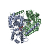
| ||||||||||||
|---|---|---|---|---|---|---|---|---|---|---|---|---|---|
| 1 |
| ||||||||||||
| Unit cell |
|
-Components
| #1: Protein | Mass: 44791.648 Da / Num. of mol.: 2 Source method: isolated from a genetically manipulated source Source: (gene. exp.) Cupriavidus necator (bacteria) / Gene: kbl, H16_B0819 / Production host: #2: Chemical |   Mass: 364.288 Da / Num. of mol.: 2 / Source method: obtained synthetically / Formula: C13H21N2O8P / Feature type: SUBJECT OF INVESTIGATION Mass: 364.288 Da / Num. of mol.: 2 / Source method: obtained synthetically / Formula: C13H21N2O8P / Feature type: SUBJECT OF INVESTIGATION#3: Water | ChemComp-HOH / |  Mass: 18.015 Da / Num. of mol.: 215 / Source method: isolated from a natural source / Formula: H2O Mass: 18.015 Da / Num. of mol.: 215 / Source method: isolated from a natural source / Formula: H2OHas ligand of interest | Y | |
|---|
-Experimental details
-Experiment
| Experiment | Method: X-RAY DIFFRACTION / Number of used crystals: 1 |
|---|
- Sample preparation
| Crystal | Density Matthews: 2.15 Å3/Da / Density % sol: 42.67 % |
|---|---|
| Crystal grow | Temperature: 295 K / Method: vapor diffusion, sitting drop Details: 0.2 M Lithium sulfate monohydrate, 0.1 M BIS-TRIS pH 6.5, 25% w/v PEG 3350, 3% w/v D-Sorbitol |
-Data collection
| Diffraction | Mean temperature: 100 K / Serial crystal experiment: N |
|---|---|
| Diffraction source | Source: SYNCHROTRON / Site: Photon Factory / Beamline: BL-5A / Wavelength: 1 Å |
| Detector | Type: DECTRIS PILATUS3 S 6M / Detector: PIXEL / Date: Dec 6, 2019 |
| Radiation | Protocol: SINGLE WAVELENGTH / Monochromatic (M) / Laue (L): M / Scattering type: x-ray |
| Radiation wavelength | Wavelength: 1 Å / Relative weight: 1 |
| Reflection | Resolution: 2.55→47.8 Å / Num. obs: 24744 / % possible obs: 99.7 % / Redundancy: 6.9 % / Biso Wilson estimate: 35.4 Å2 / CC1/2: 0.996 / Net I/σ(I): 13.1 |
| Reflection shell | Resolution: 2.55→2.64 Å / Num. unique obs: 3571 / CC1/2: 0.856 |
- Processing
| Software |
| ||||||||||||||||||||||||||||||||||||||||||||||||||||||||||||||||||||||
|---|---|---|---|---|---|---|---|---|---|---|---|---|---|---|---|---|---|---|---|---|---|---|---|---|---|---|---|---|---|---|---|---|---|---|---|---|---|---|---|---|---|---|---|---|---|---|---|---|---|---|---|---|---|---|---|---|---|---|---|---|---|---|---|---|---|---|---|---|---|---|---|
| Refinement | Method to determine structure: MOLECULAR REPLACEMENT Starting model: 1FC4 Resolution: 2.55→47.76 Å / SU ML: 0.3495 / Cross valid method: FREE R-VALUE / σ(F): 1.38 / Phase error: 25.653 Stereochemistry target values: GeoStd + Monomer Library + CDL v1.2
| ||||||||||||||||||||||||||||||||||||||||||||||||||||||||||||||||||||||
| Solvent computation | Shrinkage radii: 0.9 Å / VDW probe radii: 1.11 Å / Solvent model: FLAT BULK SOLVENT MODEL | ||||||||||||||||||||||||||||||||||||||||||||||||||||||||||||||||||||||
| Displacement parameters | Biso mean: 36.86 Å2 | ||||||||||||||||||||||||||||||||||||||||||||||||||||||||||||||||||||||
| Refinement step | Cycle: LAST / Resolution: 2.55→47.76 Å
| ||||||||||||||||||||||||||||||||||||||||||||||||||||||||||||||||||||||
| Refine LS restraints |
| ||||||||||||||||||||||||||||||||||||||||||||||||||||||||||||||||||||||
| LS refinement shell |
|
