 Movie
Movie Controller

[English] 日本語
 Yorodumi
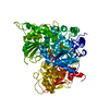
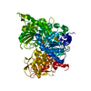
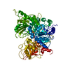
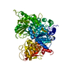
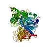
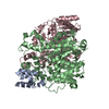
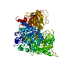
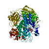
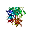
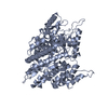
- PDB-7acn: CRYSTAL STRUCTURES OF ACONITASE WITH ISOCITRATE AND NITROISOCITRA... -
+ Open data

- Basic information
| Entry | Database: PDB / ID: 7acn | |||||||||
|---|---|---|---|---|---|---|---|---|---|---|
| Title | CRYSTAL STRUCTURES OF ACONITASE WITH ISOCITRATE AND NITROISOCITRATE BOUND | |||||||||
 Components Components | ACONITASE | |||||||||
 Keywords Keywords | LYASE(CARBON-OXYGEN) | |||||||||
| Function / homology |  Function and homology information Function and homology informationaconitate hydratase / aconitate hydratase activity / tricarboxylic acid cycle / 4 iron, 4 sulfur cluster binding / mitochondrion / metal ion binding / cytosol Similarity search - Function | |||||||||
| Biological species |  | |||||||||
| Method |  X-RAY DIFFRACTION / Resolution: 2 Å X-RAY DIFFRACTION / Resolution: 2 Å | |||||||||
 Authors Authors | Lauble, H. / Kennedy, M.C. / Beinert, H. / Stout, C.D. | |||||||||
 Citation Citation | Journal: Biochemistry / Year: 1992 Title: Crystal structures of aconitase with isocitrate and nitroisocitrate bound. Authors: Lauble, H. / Kennedy, M.C. / Beinert, H. / Stout, C.D. #1: Journal: Proc.Natl.Acad.Sci.USA / Year: 1989Title: Structure of Activated Aconitase. Formation of the (4Fe-4S) Cluster in the Crystal Authors: Robbins, A.H. / Stout, C.D. #2: Journal: Proteins / Year: 1989Title: The Structure of Aconitase Authors: Robbins, A.H. / Stout, C.D. #3: Journal: J.Biol.Chem. / Year: 1985Title: Iron-Sulfur Cluster in Aconitase. Crystallographic Evidence for a Three-Iron Center Authors: Robbins, A.H. / Stout, C.D. | |||||||||
| History |
|
- Structure visualization
| Structure viewer | Molecule: MolmilJmol/JSmol |
|---|
- Downloads & links
-Download
| PDBx/mmCIF format | 7acn.cif.gz | 164.7 KB | Display | PDBx/mmCIF format |
|---|---|---|---|---|
| PDB format | pdb7acn.ent.gz | 128.8 KB | Display | PDB format |
| PDBx/mmJSON format | 7acn.json.gz | Tree view | PDBx/mmJSON format | |
| Others |  Other downloads Other downloads |
-Validation report
| Arichive directory | https://data.pdbj.org/pub/pdb/validation_reports/ac/7acnftp://data.pdbj.org/pub/pdb/validation_reports/ac/7acn | HTTPS FTP |
|---|
-Related structure data

-Links
 PDBj
PDBj


- Assembly
| Deposited unit | 
| ||||||||
|---|---|---|---|---|---|---|---|---|---|
| 1 |
| ||||||||
| Unit cell |
| ||||||||
| Atom site foot note | 1: RESIDUE 325 IS A CIS PROLINE. / 2: SEE REMARK 5. |
-Components
| #1: Protein | Mass: 82789.047 Da / Num. of mol.: 1 Source method: isolated from a genetically manipulated source Source: (gene. exp.) |
|---|---|
| #2: Chemical | ChemComp-SF4 /   Mass: 351.640 Da / Num. of mol.: 1 / Source method: obtained synthetically / Formula: Fe4S4 Mass: 351.640 Da / Num. of mol.: 1 / Source method: obtained synthetically / Formula: Fe4S4 |
| #3: Chemical | ChemComp-ICT / 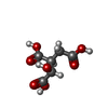  Mass: 192.124 Da / Num. of mol.: 1 / Source method: obtained synthetically / Formula: C6H8O7 Mass: 192.124 Da / Num. of mol.: 1 / Source method: obtained synthetically / Formula: C6H8O7 |
| #4: Water | ChemComp-HOH /  Mass: 18.015 Da / Num. of mol.: 338 / Source method: isolated from a natural source / Formula: H2O Mass: 18.015 Da / Num. of mol.: 338 / Source method: isolated from a natural source / Formula: H2O |
| Has protein modification | N |
-Experimental details
-Experiment
| Experiment | Method: X-RAY DIFFRACTION |
|---|
- Sample preparation
| Crystal | Density Matthews: 2.88 Å3/Da / Density % sol: 57.22 % | |||||||||||||||||||||||||
|---|---|---|---|---|---|---|---|---|---|---|---|---|---|---|---|---|---|---|---|---|---|---|---|---|---|---|
| Crystal grow | *PLUS Temperature: 22 ℃ / pH: 7 / Method: vapor diffusion / Details: referred to J.Biol.Chem. 257.9061-9063 1982 | |||||||||||||||||||||||||
| Components of the solutions | *PLUS
|
-Data collection
| Reflection | *PLUS Highest resolution: 2.1 Å / Num. all: 54710 / Num. obs: 49211 / % possible obs: 89.9 % / Num. measured all: 435630 / Rmerge F obs: 0.0683 |
|---|---|
| Reflection shell | *PLUS Highest resolution: 2.1 Å / Lowest resolution: 2.24 Å / % possible obs: 54.7 % |
- Processing
| Software |
| ||||||||||||||||||||||||||||||||||||||||||||||||||||||||||||
|---|---|---|---|---|---|---|---|---|---|---|---|---|---|---|---|---|---|---|---|---|---|---|---|---|---|---|---|---|---|---|---|---|---|---|---|---|---|---|---|---|---|---|---|---|---|---|---|---|---|---|---|---|---|---|---|---|---|---|---|---|---|
| Refinement | Rfactor Rwork: 0.179 / Rfactor obs: 0.179 / Highest resolution: 2 Å Details: RESIDUE 647 IS ARG FROM THE DNA SEQUENCE, (H. ZALKIN, PRIVATE COMMUNICATION) BUT CANNOT BE ACCOMMODATED AS SUCH IN THE STRUCTURE. IT HAS BEEN MODELED AS SER, BASED ON THE EXTENT OF THE SIDE CHAIN DENSITY. | ||||||||||||||||||||||||||||||||||||||||||||||||||||||||||||
| Refinement step | Cycle: LAST / Highest resolution: 2 Å
| ||||||||||||||||||||||||||||||||||||||||||||||||||||||||||||
| Refine LS restraints |
| ||||||||||||||||||||||||||||||||||||||||||||||||||||||||||||
| Refinement | *PLUS Highest resolution: 2.1 Å / Rfactor obs: 0.161 / Lowest resolution: 8 Å / Rfactor Rwork: 0.161 | ||||||||||||||||||||||||||||||||||||||||||||||||||||||||||||
| Solvent computation | *PLUS | ||||||||||||||||||||||||||||||||||||||||||||||||||||||||||||
| Displacement parameters | *PLUS | ||||||||||||||||||||||||||||||||||||||||||||||||||||||||||||
| Refine LS restraints | *PLUS Type: x_angle_d / Dev ideal: 3.09 |
