National Institutes of Health/National Institute of General Medical Sciences (NIH/NIGMS)
1R01GM108921
United States
National Institutes of Health/National Institute of General Medical Sciences (NIH/NIGMS)
3R01GM108921-03S1
United States
American Heart Association
17POST33671152
United States
Citation
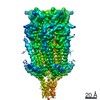
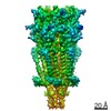
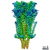
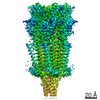
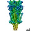
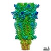
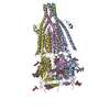
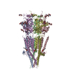
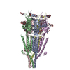
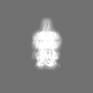
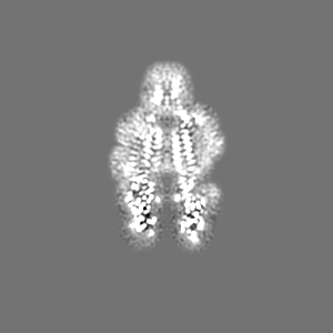
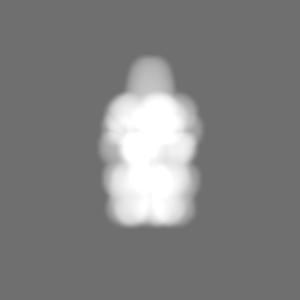
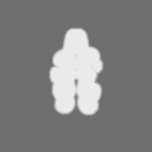
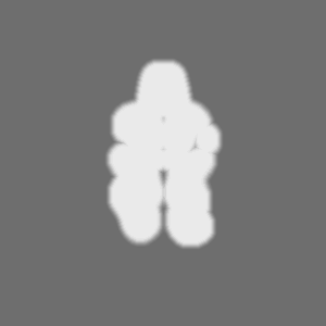
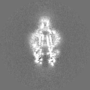
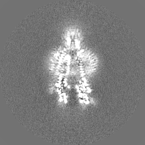
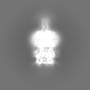
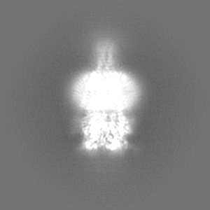
Journal: Nature / Year: 2018 Title: Cryo-EM reveals two distinct serotonin-bound conformations of full-length 5-HT receptor. Authors: Sandip Basak / Yvonne Gicheru / Shanlin Rao / Mark S P Sansom / Sudha Chakrapani / Abstract: The 5-HT serotonin receptor, a cationic pentameric ligand-gated ion channel (pLGIC), is the clinical target for management of nausea and vomiting associated with radiation and chemotherapies. Upon ...The 5-HT serotonin receptor, a cationic pentameric ligand-gated ion channel (pLGIC), is the clinical target for management of nausea and vomiting associated with radiation and chemotherapies. Upon binding, serotonin induces a global conformational change that encompasses the ligand-binding extracellular domain (ECD), the transmembrane domain (TMD) and the intracellular domain (ICD), the molecular details of which are unclear. Here we present two serotonin-bound structures of the full-length 5-HT receptor in distinct conformations at 3.32 Å and 3.89 Å resolution that reveal the mechanism underlying channel activation. In comparison to the apo 5-HT receptor, serotonin-bound states underwent a large twisting motion in the ECD and TMD, leading to the opening of a 165 Å permeation pathway. Notably, this motion results in the creation of lateral portals for ion permeation at the interface of the TMD and ICD. Combined with molecular dynamics simulations, these structures provide novel insights into conformational coupling across domains and functional modulation.
History
Deposition
May 17, 2018
-
Header (metadata) release
May 30, 2018
-
Map release
Nov 7, 2018
-
Update
Nov 20, 2024
-
Current status
Nov 20, 2024
Processing site: RCSB / Status: Released
-
Structure visualization
Movie
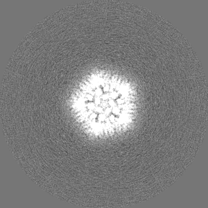
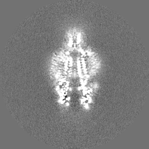
Surface view with section colored by density value
Model: Quantifoil R1.2/1.3 / Material: COPPER / Mesh: 300 / Support film - Material: CARBON / Support film - topology: HOLEY / Pretreatment - Type: GLOW DISCHARGE / Pretreatment - Time: 15 sec.
Vitrification
Cryogen name: ETHANE / Chamber humidity: 100 % / Chamber temperature: 277 K / Instrument: FEI VITROBOT MARK I / Details: blot for 2.5 seconds.
-
Electron microscopy
Microscope
FEI TITAN KRIOS
Image recording
Film or detector model: GATAN K2 SUMMIT (4k x 4k) / Detector mode: SUPER-RESOLUTION / Digitization - Frames/image: 2-39 / Number real images: 2810 / Average electron dose: 40.0 e/Å2
Electron beam
Acceleration voltage: 300 kV / Electron source: FIELD EMISSION GUN
In the structure databanks used in Yorodumi, some data are registered as the other names, "COVID-19 virus" and "2019-nCoV". Here are the details of the virus and the list of structure data.
Jan 31, 2019. EMDB accession codes are about to change! (news from PDBe EMDB page)
EMDB accession codes are about to change! (news from PDBe EMDB page)
The allocation of 4 digits for EMDB accession codes will soon come to an end. Whilst these codes will remain in use, new EMDB accession codes will include an additional digit and will expand incrementally as the available range of codes is exhausted. The current 4-digit format prefixed with “EMD-” (i.e. EMD-XXXX) will advance to a 5-digit format (i.e. EMD-XXXXX), and so on. It is currently estimated that the 4-digit codes will be depleted around Spring 2019, at which point the 5-digit format will come into force.
The EM Navigator/Yorodumi systems omit the EMD- prefix.
Related info.:Q: What is EMD? / ID/Accession-code notation in Yorodumi/EM Navigator
Yorodumi is a browser for structure data from EMDB, PDB, SASBDB, etc.
This page is also the successor to EM Navigator detail page, and also detail information page/front-end page for Omokage search.
The word "yorodu" (or yorozu) is an old Japanese word meaning "ten thousand". "mi" (miru) is to see.
Related info.:EMDB / PDB / SASBDB / Comparison of 3 databanks / Yorodumi Search / Aug 31, 2016. New EM Navigator & Yorodumi / Yorodumi Papers / Jmol/JSmol / Function and homology information / Changes in new EM Navigator and Yorodumi
 Movie
Movie Controller

 Yorodumi
 Open data
 Basic information
 Map data
 Sample
 Keywords
 Function and homology information
 Authors
 United States, 3 items
  Citation
 Structure visualization
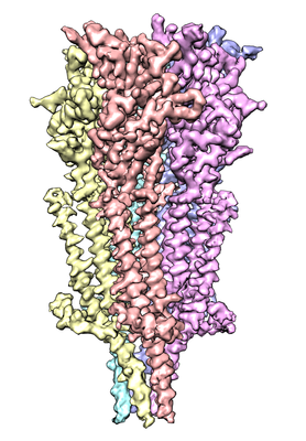
 Downloads & links
 emd_7882.png
 http://ftp.pdbj.org/pub/emdb/structures/EMD-7882


 Z (Sec.)
 Y (Row.)
 X (Col.)


 Sample components
 Spodoptera frugiperda (fall armyworm)

 Processing
 Electron microscopy
 FIELD EMISSION GUN