[English] 日本語
 Yorodumi
- PDB-6zvs: C12 symmetry: Bacterial Vipp1 and PspA are members of the ancient... -
+ Open data
- Basic information
| Entry | Database: PDB / ID: 6zvs | ||||||
|---|---|---|---|---|---|---|---|
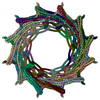
| Title | C12 symmetry: Bacterial Vipp1 and PspA are members of the ancient ESCRT-III membrane-remodeling superfamily. | ||||||
 Components Components | Vipp1 | ||||||
 Keywords Keywords | LIPID BINDING PROTEIN / membrane remodelling | ||||||
| Function / homology | PspA/IM30 / PspA/IM30 family / lipid binding / plasma membrane / Membrane-associated protein Vipp1 Function and homology information Function and homology information | ||||||
| Biological species |  Nostoc punctiforme (bacteria) Nostoc punctiforme (bacteria) | ||||||
| Method | ELECTRON MICROSCOPY / single particle reconstruction / cryo EM / Resolution: 7.2 Å | ||||||
 Authors Authors | Liu, J. / Tassinari, M. / Souza, D.P. / Naskar, S. / Noel, J.K. / Bohuszewicz, O. / Buck, M. / Williams, T.A. / Baum, B. / Low, H.H. | ||||||
| Funding support |  United Kingdom, 1items United Kingdom, 1items
| ||||||
 Citation Citation |  Journal: Cell / Year: 2021 Journal: Cell / Year: 2021Title: Bacterial Vipp1 and PspA are members of the ancient ESCRT-III membrane-remodeling superfamily. Authors: Jiwei Liu / Matteo Tassinari / Diorge P Souza / Souvik Naskar / Jeffrey K Noel / Olga Bohuszewicz / Martin Buck / Tom A Williams / Buzz Baum / Harry H Low /   Abstract: Membrane remodeling and repair are essential for all cells. Proteins that perform these functions include Vipp1/IM30 in photosynthetic plastids, PspA in bacteria, and ESCRT-III in eukaryotes. Here, ...Membrane remodeling and repair are essential for all cells. Proteins that perform these functions include Vipp1/IM30 in photosynthetic plastids, PspA in bacteria, and ESCRT-III in eukaryotes. Here, using a combination of evolutionary and structural analyses, we show that these protein families are homologous and share a common ancient evolutionary origin that likely predates the last universal common ancestor. This homology is evident in cryo-electron microscopy structures of Vipp1 rings from the cyanobacterium Nostoc punctiforme presented over a range of symmetries. Each ring is assembled from rungs that stack and progressively tilt to form dome-shaped curvature. Assembly is facilitated by hinges in the Vipp1 monomer, similar to those in ESCRT-III proteins, which allow the formation of flexible polymers. Rings have an inner lumen that is able to bind and deform membranes. Collectively, these data suggest conserved mechanistic principles that underlie Vipp1, PspA, and ESCRT-III-dependent membrane remodeling across all domains of life. | ||||||
| History |
|
- Structure visualization
| Movie |
 Movie viewer Movie viewer |
|---|---|
| Structure viewer | Molecule:  Molmil Molmil Jmol/JSmol Jmol/JSmol |
- Downloads & links
- Download
| PDBx/mmCIF format |  6zvs.cif.gz 6zvs.cif.gz | 1.8 MB | Display |  PDBx/mmCIF format PDBx/mmCIF format |
|---|---|---|---|---|
| PDB format |  pdb6zvs.ent.gz pdb6zvs.ent.gz | Display |  PDB format PDB format | |
| PDBx/mmJSON format |  6zvs.json.gz 6zvs.json.gz | Tree view |  PDBx/mmJSON format PDBx/mmJSON format | |
| Others |  Other downloads Other downloads |
-Validation report
| Summary document |  6zvs_validation.pdf.gz 6zvs_validation.pdf.gz | 1.2 MB | Display |  wwPDB validaton report wwPDB validaton report |
|---|---|---|---|---|
| Full document |  6zvs_full_validation.pdf.gz 6zvs_full_validation.pdf.gz | 1.2 MB | Display | |
| Data in XML |  6zvs_validation.xml.gz 6zvs_validation.xml.gz | 212.2 KB | Display | |
| Data in CIF |  6zvs_validation.cif.gz 6zvs_validation.cif.gz | 380.4 KB | Display | |
| Arichive directory |  https://data.pdbj.org/pub/pdb/validation_reports/zv/6zvs https://data.pdbj.org/pub/pdb/validation_reports/zv/6zvs ftp://data.pdbj.org/pub/pdb/validation_reports/zv/6zvs ftp://data.pdbj.org/pub/pdb/validation_reports/zv/6zvs | HTTPS FTP |
-Related structure data
| Related structure data |  11469MC  6zvrC  6zvtC  6zw4C  6zw5C  6zw6C  6zw7C C: citing same article ( M: map data used to model this data |
|---|---|
| Similar structure data |
- Links
Links
- Assembly
| Deposited unit | 
|
|---|---|
| 1 |
|
- Components
| #1: Protein | Mass: 28745.461 Da / Num. of mol.: 72 Source method: isolated from a genetically manipulated source Source: (gene. exp.)  Nostoc punctiforme (bacteria) / Production host: Nostoc punctiforme (bacteria) / Production host:  |
|---|
-Experimental details
-Experiment
| Experiment | Method: ELECTRON MICROSCOPY |
|---|---|
| EM experiment | Aggregation state: PARTICLE / 3D reconstruction method: single particle reconstruction |
- Sample preparation
| Component | Name: vipp1 c12 ring / Type: COMPLEX / Entity ID: all / Source: RECOMBINANT |
|---|---|
| Source (natural) | Organism:  Nostoc punctiforme (bacteria) Nostoc punctiforme (bacteria) |
| Source (recombinant) | Organism:  |
| Buffer solution | pH: 8.4 |
| Specimen | Embedding applied: NO / Shadowing applied: NO / Staining applied: NO / Vitrification applied: YES |
| Vitrification | Cryogen name: ETHANE |
- Electron microscopy imaging
| Experimental equipment |  Model: Titan Krios / Image courtesy: FEI Company |
|---|---|
| Microscopy | Model: FEI TITAN KRIOS |
| Electron gun | Electron source:  FIELD EMISSION GUN / Accelerating voltage: 300 kV / Illumination mode: FLOOD BEAM FIELD EMISSION GUN / Accelerating voltage: 300 kV / Illumination mode: FLOOD BEAM |
| Electron lens | Mode: BRIGHT FIELD |
| Image recording | Electron dose: 1.5 e/Å2 / Film or detector model: GATAN K2 SUMMIT (4k x 4k) |
- Processing
| Software |
| ||||||||||||||||||||||||
|---|---|---|---|---|---|---|---|---|---|---|---|---|---|---|---|---|---|---|---|---|---|---|---|---|---|
| EM software | Name: RELION / Category: 3D reconstruction | ||||||||||||||||||||||||
| CTF correction | Type: NONE | ||||||||||||||||||||||||
| 3D reconstruction | Resolution: 7.2 Å / Resolution method: FSC 0.143 CUT-OFF / Num. of particles: 7433 / Symmetry type: POINT | ||||||||||||||||||||||||
| Refinement | Cross valid method: NONE Stereochemistry target values: GeoStd + Monomer Library + CDL v1.2 | ||||||||||||||||||||||||
| Displacement parameters | Biso mean: 255.97 Å2 | ||||||||||||||||||||||||
| Refine LS restraints |
|
 Movie
Movie Controller


 PDBj
PDBj